3.22 The Labeled Cell Gallery: What Our Station Reveals
use our station’s images to build a labeled cell gallery that shows living things are made of cells and explains what each visible cell part does.
- I can analyze my data to decide if the evidence shows that cellular composition is a feature that distinguishes living things.
- I can use my model to describe how each part of the cell, like the nucleus, chloroplasts, mitochondria, cell membrane, and cell wall, helps the cell do its job.
- I can explain how the structure of the cell membrane or cell wall relates to their function and affects how the cell and its parts work together.
- I can use my model to compare plant and animal cells by showing that plant cells have both a cell wall and a cell membrane, while animal cells only have a cell membrane.
- I can use my model to explain that plant cells have chloroplasts for photosynthesis, but animal cells do not.
Phase 4: Build
Warm-Up ~5 min
Do Now. A visitor to the clinic asks, “Is a drop of pond water alive?” What would you need to see in a picture to answer yes for at least one thing in the drop?
What is the smallest unit that can be alive?
Instruction ~10 min
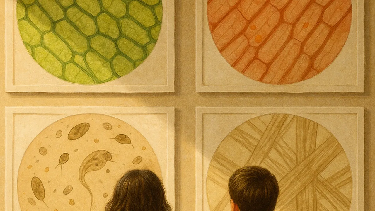
The gallery is our evidence. Each team photographs four specimens with its station: stained onion skin, an elodea leaf, a drop of pond water and a drop of yeast mixed with warm water. A prepared slide of animal tissue gives us a fifth image to compare. Each photo gets labels and a caption.
What each part does. The cell membrane is a thin boundary around every cell that controls what goes in and out. Plant cells, yeast and many algae also have a stiff cell wall outside the membrane that supports the cell and keeps its shape. The nucleus holds the cell’s instructions and controls its activities. Chloroplasts in green plant cells use light to make food. Mitochondria release energy from food for the cell to use. They are in almost all of these cells, but they are too small to see clearly with most school microscopes. The parts work together: food from chloroplasts becomes usable energy in mitochondria, and the membrane lets in what the cell needs.
Worked example 1: an elodea caption. “Elodea leaf, about 100×. Rows of box-shaped cells. Each has a cell wall (thick outline) and many green chloroplasts. The chloroplasts make food from light. The nucleus is hard to see because chloroplasts crowd the view.”
Worked example 2: a pond-water caption. “Pond water, about 100×. One moving organism made of a single cell, plus a chain of green cells. The single-celled organism is a whole living thing. The paper fibers we tested in 3.19 had no living cells.” This is how the gallery shows that living things are made of one cell or many cells.
| Specimen | One cell or many? | Cell wall? | Nucleus seen? | Chloroplasts? | Caption written? |
|---|---|---|---|---|---|
| Onion + iodine | |||||
| Elodea | |||||
| Pond water | |||||
| Yeast | |||||
| Prepared animal tissue |
Guided Exercises ~15 min
Photograph each specimen, add labels, then answer the block that goes with it.
Independent Practice ~10 min
Finish your part of the gallery.
Draw and label a model of an elodea cell and an animal cell side by side. Include nucleus, membrane, mitochondria, and wall and chloroplasts where they belong.
Write a two-sentence gallery caption for a family visitor. Explain what the cell is, and how one part helps the whole cell stay alive.
Closure ~5 min
The gallery is taking shape. Next class, other teams critique our stations and we revise.
Exit ticket: what is the purpose of the cell gallery as science evidence? Use the words cell and living.
How confident are you that you can I can use a model to describe how each cell part helps the cell do its job.
Teacher notes
Materials: onion, iodine, elodea, pond water collected by the teacher, dry yeast with warm water and a pinch of sugar, a prepared animal tissue slide, slides, cover slips, droppers, goggles, gloves, labeling app or printouts.
Setup: collect pond water a day ahead from a safe spot and keep it in a lidded jar near light. No cheek swabs or blood. Yeast is a single-celled fungus with a cell wall; it is neither plant nor animal.
Listen for: “animal cells have no nucleus”; “the wall replaces the membrane” (plant cells have both); “we cannot see it, so it is not there.”
Support / stretch: give a word bank and pre-printed images to label. Stretch: compare cell counts across the view in onion vs elodea and explain the size difference.