3.06 First Look: Onion Skin, Elodea and Pond Water
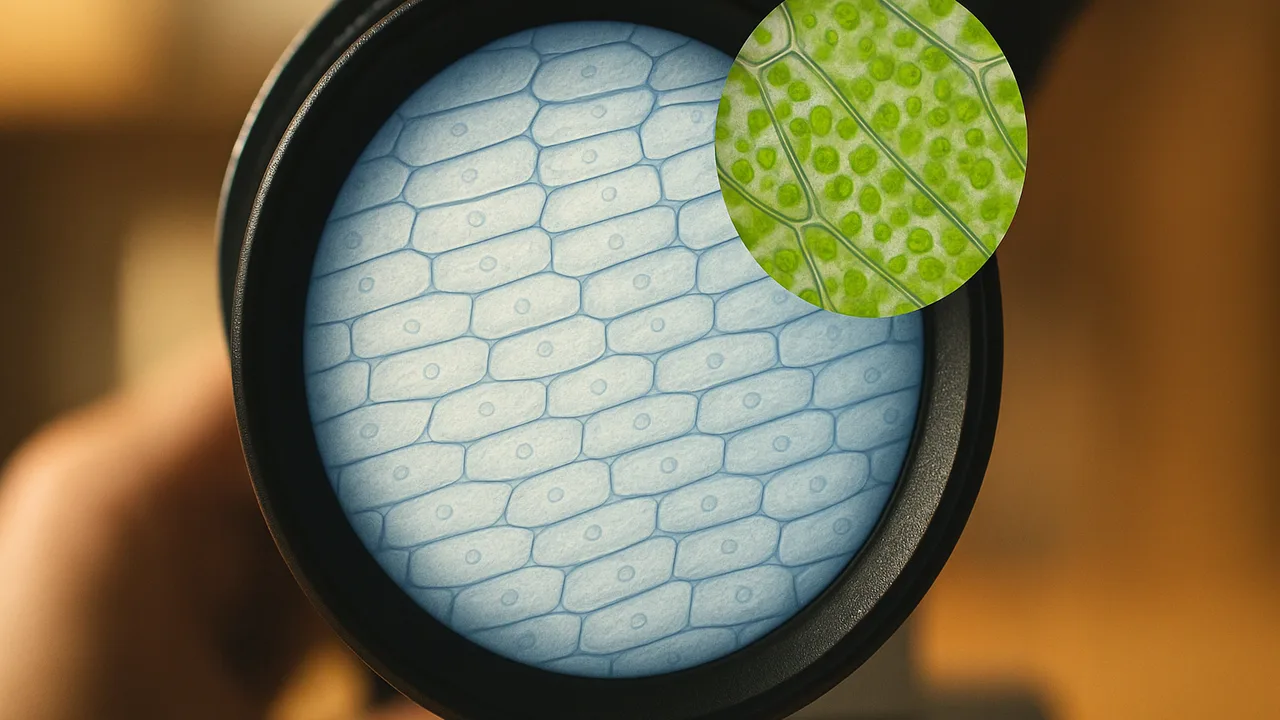
use a microscope to observe and record evidence of cells in onion skin, elodea and pond water, and explain why the investigation needs magnification.
- I can collect and record data about the cellular makeup of living organisms.
- I can identify the tools I use to observe cells at different magnifications and explain why different tools are needed to see cells at different scales.
- I can explain the purpose of my investigation: to show that all living things are made of cells, and that the cell is the smallest unit that can be alive.
Phase 2: Investigate
Warm-Up ~5 min
Do Now. Peel the thin, see-through layer from the inside of an onion piece. Hold it up to the light and look with a hand lens. Then predict.
At 100× under a microscope, what will this onion layer look like?
Instruction ~10 min
Purpose of our investigation. We are testing the claim that all living things are made of cells and that the cell is the smallest unit that can be alive. Today you collect the first evidence.
Using the microscope. Start on the lowest power objective. Use the coarse focus knob only at low power. Center what you want to see, then switch to a higher objective and use only the fine focus knob. Draw what you see in a circle and write the magnification beside it.
Three stations.
- Onion skin: a thin layer in a drop of water. Add one drop of iodine stain at the edge of the cover slip.
- Elodea: one young leaf of this water plant in a drop of water.
- Pond water: one drop from the bottom of the jar, where life gathers.
| Sample | Magnification | Cells seen? (yes / no) | Shape and arrangement | Parts visible inside | Moving? | Sketch number |
|---|---|---|---|---|---|---|
| Onion skin | ||||||
| Elodea leaf | ||||||
| Pond water |
Worked example 1: recording an observation. Jamal’s notebook: “Onion, 100×: rows of long, brick-shaped cells. Clear walls between them. One dark round spot in most cells after iodine. No movement.” He recorded magnification, shape, arrangement and parts. He did not write “cool” or “weird.”
Worked example 2: choosing the right tool. Ava could see the onion layer with her eyes, but no cells. At 5× with a hand lens, she saw faint lines. At 100× she saw whole cells. At 400×, small green ovals inside elodea cells became clear, and some drifted slowly around the edges. Each jump in magnification showed a smaller level of detail. Without engineered lenses, none of this evidence would exist.
Guided Exercises ~15 min
Answer with your data table and sketches beside you.
Independent Practice ~10 min
Sketch with care.
Draw one cell from each sample at the magnification you used. Label the magnification and any parts you saw. Under each drawing, write one sentence about how it supports or does not support the claim that living things are made of cells.
How confident are you that you can I can use a microscope to collect and record data about cells.
Closure ~5 min
Some pond creatures were whole cells swimming on their own. Next time, we compare one-celled and many-celled living things, and plant and animal cells.
Exit ticket: name the tool you would use to see (a) a whole onion, (b) faint lines in onion skin, and (c) chloroplasts in elodea. Explain why each scale needs a different tool.
Teacher notes
Materials (per team): compound microscope, slides, cover slips, droppers, onion, elodea (from an aquarium or science supplier), pond water (or a hay infusion started a week ahead), iodine stain in a dropper bottle, goggles, gloves, paper towels.
Setup: run three 10-minute stations. Pre-make one backup slide of each. If you have a microscope camera, project one view for the class.
Listen for: “bubbles are cells.” Air bubbles are round, dark-edged and empty; teach students to spot them. Chloroplasts in elodea often move with the cell’s fluid; students may see it at 400×. Mitochondria are too small to see clearly with school microscopes; do not claim they are visible.
Support / stretch: pre-made slides for students who struggle with fine motor work. Stretch: estimate how many onion cells span the field of view at 100×.