3.02 Mapping the Microscope Station System
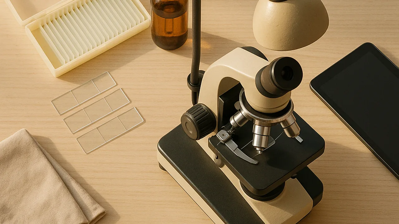
Seeing the Invisible: A Digital Microscope Station
How can light, lenses and digital signals let us see and share things far too small for our eyes, and what do we find when we look?
The mystery: A drop of pond water looks clear. Under a microscope it is full of moving living things. A phone camera clipped to a cheap lens can send that picture across the world in a second.
map the parts, users and boundary of the microscope station system and explain how better tools have let scientists discover cells.
- I can identify the system where the problem exists, including its main parts, how they relate, and what is inside or outside the system.
- I can explain who needs the problem solved, what needs must be met, and which scientific issues are important.
- I can analyze how possible solutions might affect people and the environment, and decide which issues and parts of the system matter most.
- I can explain how the tools I use in my investigation show that science depends on engineering advances.
Phase 1: Spark
Warm-Up ~5 min
Do Now. List every part you think a digital microscope station needs, from the sample to the family’s phone at home. Then answer the prediction.
Which came first: the idea that living things are made of cells, or the tool that let people see cells?
Instruction ~10 min
Thinking in systems. A system is a group of parts that work together. Engineers draw a system map to see every part, how the parts connect and where the system’s boundary is. Things inside the boundary are parts we can design. Things outside still affect the system.
Worked example 1: mapping our station. Mei’s team listed these parts inside the boundary: the sample on a slide, a light source, one or more lenses, a phone or small camera, a screen and a way to send images. Outside the boundary they listed the clinic staff, students, families, the room’s lighting, the internet connection and the budget. Arrows showed light going from the lamp, through the sample, through the lens and into the camera.
Worked example 2: who needs what? Jamal’s team asked what each user needs. Clinic staff need a station that is quick to set up and easy to clean. Young children need a big, bright image. Families at home need images they can open on any phone. Scientific issues include how much to magnify and how sharp the image must be. Then the team starred the parts that matter most: the lens and the camera.
Science depends on engineering. In 1665, Robert Hooke used an early microscope to look at a thin slice of cork. He saw tiny boxes and called them “cells.” In the 1670s, Antonie van Leeuwenhoek ground his own tiny, powerful lenses. He was among the first people to see living one-celled organisms in water. Better lenses led to better evidence. Today digital cameras and computers let scientists record and share cell images around the world. Our station is one more step in that story.
Guided Exercises ~15 min
Build the class system map together, card by card.
Independent Practice ~10 min
Draw your own system map in your notebook.
Draw a system map of the microscope station. Show at least five parts inside the boundary and three things outside it. Star the two parts that matter most, and explain why.
How confident are you that you can I can map a system and explain who needs the problem solved.
Closure ~5 min
Light is the key input to our whole system. Next time, the Investigate phase starts with the simplest wave of all: one on a long spring.
Exit ticket: name two users of the station and one need each has. Then explain one way a better tool helped scientists learn about cells.
Teacher notes
Materials: chart paper, markers, sticky notes, an old phone and a hand lens to pass around, images of an early microscope (many museum websites share them).
Setup: keep the class system map posted all unit. Teams add to it after each investigation.
Listen for: students who leave people out of the system. Remind them that users shape every design choice. Keep the history general: Hooke’s cork observation (1665) and van Leeuwenhoek’s single-lens microscopes (1670s) are well documented; avoid other specific claims.
Support / stretch: give a partly drawn map with blank boxes. Stretch: add a second boundary for the families’ homes and show what crosses it.