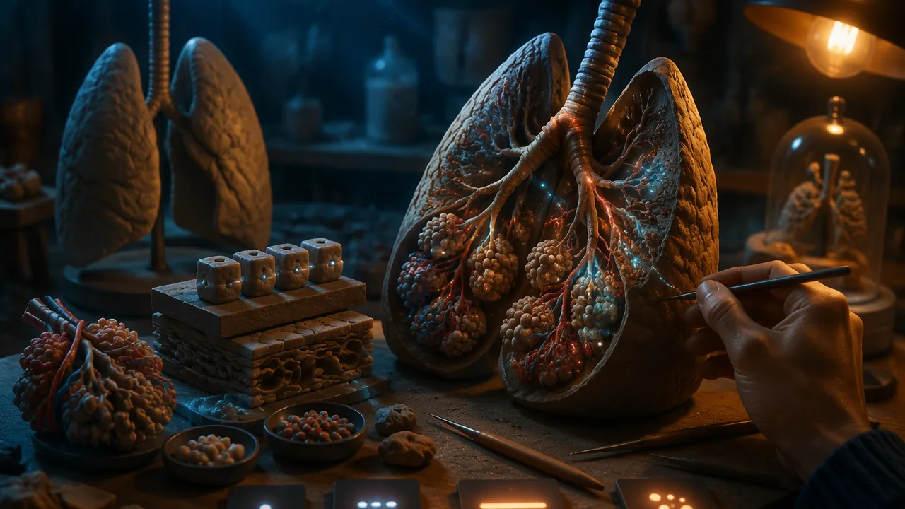
describe the levels of organization and the main structures and functions of the eleven organ systems, and sculpt a labeled model that shows how one system’s structure serves its function.
Instruction
OpenStax Anatomy and Physiology 2e (section 1.2) describes the body at six levels of organization: chemical (atoms and molecules), cellular, tissue (groups of similar cells working together), organ (two or more tissue types forming a structure with a function), organ system (organs working together) and organism. Your model sits at the organ-system level, but it should hint at the levels below.
The eleven organ systems. OpenStax groups the body into eleven systems. The integumentary system (skin, hair, nails) protects, regulates temperature and senses. The skeletal system (bones, cartilage, ligaments) supports, protects organs, stores minerals and makes blood cells in bone marrow. The muscular system (skeletal muscles and tendons) moves the body and generates heat. The nervous system (brain, spinal cord, nerves) receives, processes and sends signals. The endocrine system (glands such as the pituitary, thyroid, pancreas and adrenal glands) sends chemical signals called hormones through the blood. The cardiovascular system (heart and blood vessels) pumps and carries blood, delivering oxygen and nutrients and removing wastes. The lymphatic system (lymph nodes, vessels, spleen, thymus) returns fluid to the blood and houses immune cells. The respiratory system (nasal passages, trachea, lungs) exchanges oxygen and carbon dioxide. The digestive system (mouth, esophagus, stomach, intestines, liver, pancreas) breaks down and absorbs food. The urinary system (kidneys, ureters, bladder, urethra) filters blood and removes wastes in urine while regulating water and salt. The reproductive systems produce sex cells and hormones and, in females, support a developing fetus.
Structure serves function. A good model makes one structure-function relationship obvious. The lungs’ millions of tiny air sacs (alveoli) give an enormous surface area for gas exchange; a model might show one branch ending in a cluster of small spheres. The small intestine’s folded lining and finger-like villi increase surface area for absorption. The heart’s thick left ventricle wall pumps blood to the whole body. Choose the relationship first, then design the model to show it.
Modeling decisions. Decide on scale (life-size, enlarged or reduced) and say which. Decide what to simplify: you cannot sculpt every nerve, so show the pathway. Use color as code, with a key. Use an armature (a wire or rolled-foil skeleton inside the clay) for anything that must stand up. Consider an exploded or cutaway form: a heart cut open shows the chambers.
How clinicians examine a system. This course asks you to know the basic physical examination. Clinicians use four classic techniques: inspection (looking), palpation (feeling with the hands), percussion (tapping to hear whether a region is hollow or solid) and auscultation (listening, usually with a stethoscope, to the heart, lungs or bowel). Your key card should name which techniques a clinician would use for your system, as a bridge to the next lesson.
Craft tips. Keep clay pieces thinner than about 2 cm so they dry evenly. Join pieces by scoring both surfaces and pressing them together with a little water. Label with small flags on toothpicks or with a separate numbered key.
📖 Read for this lesson
The reading opens over the lesson; close it to come back to exactly where you were. The checks below draw on it.
Studio: Clay organ systems
Question. How can a hand-built model make one structure-function relationship of an organ system clear to a viewer who has never studied anatomy?
Materials
- Air-dry clay or homemade salt dough (about 500 g)
- Aluminum foil and pipe cleaners or soft craft wire for armatures
- Toothpicks, paper flags and a fine marker for labels
- Acrylic or tempera paint and brushes (optional)
- Paper plates or card bases, and plastic modeling tools or craft sticks
- OpenStax Anatomy and Physiology 2e figures for the system you choose (the reading for this lesson describes all eleven)
- Salt dough and some modeling doughs contain wheat; if you have a wheat or gluten allergy, use gluten-free air-dry clay and wear gloves if needed. Check the package label before you start.
- Bend the ends of wire back on themselves or cover them with tape so they cannot scratch or puncture skin. Have an adult present if you cut wire with pliers or use a craft knife.
- Use water-based paints only; wash hands after handling clay and paint and never eat at your work area.
- Wipe clay dust with a damp cloth rather than sweeping, to avoid breathing dust.
Procedure
- Choose one system, draw it from the OpenStax figure and circle the one structure-function relationship your model will make obvious.
- Sketch two model designs (for example, whole-system and cutaway of one organ) and choose one. Note the scale you will use.
- Build an armature from foil or wire for any part that must stand up, and cover it with clay in layers no thicker than about 2 cm.
- Score and join separate pieces with a little water. Add surface texture that shows function, such as folds, branches or chambers.
- Let the model dry, paint it with a color code if you wish, and add labels on flags or a numbered key.
- Write a key card: system, scale, the structure-function relationship shown, what you simplified, and which examination techniques a clinician would use for this system.
- Photograph the model from three angles in good light for your atlas panel.
Data
| System | Scale | Structure-function relationship shown | What was simplified | Examination techniques |
|---|---|---|---|---|
Analysis
- Put the model away for a day. Then cover your key card and look at the model as a first-time viewer: what would they notice first, and could they say what function it shows? If you like, show it to a family member or other adult who has not studied your system and write down what they say first, without explaining.
- Which modeling decision (scale, simplification, color, view) did the most to make the function clear? Which would you change?
- Compare your model with the OpenStax figure. Name one thing the model shows better and one thing the figure shows better.
- How would you turn this model into a digital or interactive version later in the course?
If you cannot do this studio in person: If you have no clay, build the model from household materials such as foil, paper, pasta, string or play dough, or make it digitally in Blender (free, no account needed), and photograph it from three angles.
Formative check
Work through these before moving on. They are not graded — they tell you whether the standard below has landed yet.
Which system includes the pituitary, thyroid and adrenal glands and signals through hormones in the blood?
The four classic physical examination techniques are inspection, , percussion and auscultation.
Which structure-function relationship would a model of the respiratory system best show with a cluster of tiny spheres at the end of each branch?
Integumentary
Tap to revealSkin, hair, nails: protection, temperature, sensation
Lymphatic
Tap to revealReturns fluid to blood; houses immune cells
Urinary
Tap to revealFilters blood; removes wastes; regulates water and salt
Skeletal
Tap to revealSupport, protection, mineral storage, blood-cell production
Cardiovascular
Tap to revealHeart and vessels carry oxygen, nutrients and wastes
How confident are you that you can name the eleven organ systems and sculpt a model that makes one structure-function relationship clear?
Practice
Work these on paper or in your notebook, then open Check your answer. Aim for all of Fluency and Application; try at least one Challenge.
Printable version: this unit’s practice workbook (PDF)
Fluency
Build speed and accuracy with the core skill.
- List the six levels of organization from smallest to largest.
Check your answer
Answer: Chemical, cellular, tissue, organ, organ system, organism.OpenStax Anatomy and Physiology 2e, section 1.2. An organ is two or more tissue types forming a structure with a function. - Name the organ system: (a) kidneys, ureters, bladder; (b) lymph nodes, spleen, thymus; (c) skin, hair, nails; (d) brain, spinal cord, nerves.
Check your answer
Answer: (a) urinary; (b) lymphatic; (c) integumentary; (d) nervous.The eleven systems in OpenStax: integumentary, skeletal, muscular, nervous, endocrine, cardiovascular, lymphatic, respiratory, digestive, urinary, reproductive. - Name the four classic physical examination techniques and what each one involves.
Check your answer
Answer: Inspection (looking), palpation (feeling with the hands), percussion (tapping to hear whether a region is hollow or solid), auscultation (listening, usually with a stethoscope).Your key card names which techniques a clinician would use for your system.
Application
Use the skill in context. Show your reasoning.
- For each structure, state the function it serves and one way a model could make that visible: (a) alveoli; (b) villi of the small intestine; (c) the thick left ventricle wall.
Check your answer
Answer: (a) Huge surface area for gas exchange; end each branch in a cluster of small spheres. (b) Increased surface area for absorption; sculpt a folded lining with many finger-like projections. (c) Pumping blood to the whole body; a cutaway heart with the left wall visibly thicker than the right.Choose the relationship first, then design the model to show it. - A heart model is enlarged 3 times. If a heart is drawn from a reference as 12 cm tall, how tall is the model? The lesson says keep clay thinner than about 2 cm; why might a solid 36 cm heart be a problem?
Check your answer
Answer: 36 cm. A solid piece that large would be far thicker than 2 cm and would not dry evenly, so build it over an armature or as a hollow or cutaway form.12 x 3 = 36. Thin layers over a foil armature dry evenly; a cutaway also shows the chambers. - Write a key card for a clay model of the respiratory system that shows the alveoli. Include: system, scale, the structure-function relationship, what you simplified, and examination technique(s).
Check your answer
Answer: Model: System: respiratory. Scale: enlarged (alveoli shown far larger than life). Relationship: millions of tiny alveoli give an enormous surface area for gas exchange. Simplified: only one branch shown in detail; blood vessels and most branches left out. Examination: auscultation (listening to the lungs), plus inspection.A strong card states the scale honestly and names what was left out. Listening to the lungs is the technique the lesson names for lungs. - Match each function to its system: (a) sends chemical signals called hormones through the blood; (b) stores minerals and makes blood cells in bone marrow; (c) breaks down and absorbs food; (d) filters blood and regulates water and salt.
Check your answer
Answer: (a) endocrine; (b) skeletal; (c) digestive; (d) urinary.From the lesson’s descriptions of the eleven systems.
Review
Keep earlier skills sharp.
- Review 3.1: Your model of the heart is cut open to show the chambers. Which view type is that, and which alternative view would separate the parts slightly to show how they fit?
Check your answer
Answer: A cutaway; the alternative is an exploded view.Both are named in 3.1 and recommended in 3.2’s modeling decisions.
Eleven Organ Systems: A Modeling Sheet with One Relationship Each, Scale Arithmetic and a Worked Key Card
SLS original modeling sheet; structures and functions restate the mini-lecture’s level of detail and no textbook text is used; sources for any card statement to be added by the student from MedlinePlus or NIH; prepared Sept. 26, 2026 · SLS original
Why this reading. The lab tells you to draw your system from a textbook figure and to circle one structure-function relationship, and that figure cannot be hosted here. This SLS original sheet supplies what you need instead: for each of the eleven systems, its main structures and functions (the same ones the mini-lecture names), one relationship that makes a good model, and how to build it; plus a worked key card and the scale arithmetic. It contains no text from any textbook. If you also have a figure from a textbook or a federal health site, use it to check your drawing, not as the only source.
Eleven systems, one relationship each
| System | Main structures | Main functions | A relationship worth modeling | How to model it |
|---|---|---|---|---|
| Integumentary | Skin, hair, nails | Protection, temperature control, sensation | Layers: a thin outer layer over a thicker inner layer | Two colors of clay, layered, with a hair or gland as a pin |
| Skeletal | Bones, cartilage, ligaments | Support, protection, mineral storage, making blood cells in marrow | A hollow-looking long bone with a marrow space | Cut a foil-and-clay long bone lengthwise to show the marrow cavity |
| Muscular | Skeletal muscles, tendons | Movement, heat | A muscle pulls a bone through a tendon | Two clay bones joined with wire, a muscle strap and a tendon |
| Nervous | Brain, spinal cord, nerves | Receive, process and send signals | Branching: one cord and many nerves reaching out | Wire branches from a central cord |
| Endocrine | Glands: pituitary, thyroid, pancreas, adrenals | Hormone signals carried in blood | A gland releases into a vessel, not through a duct to a surface | Small colored beads next to a blood-vessel tube |
| Cardiovascular | Heart and vessels | Carry oxygen and nutrients; remove wastes | A pump with one-way flow | A two-sided pump with arrows showing the direction of flow |
| Lymphatic | Vessels, nodes, spleen, thymus | Return fluid to blood; house immune cells | Nodes as collection points along vessels | Beads (nodes) on a string (vessels) |
| Respiratory | Nasal passages, trachea, lungs | Exchange oxygen and carbon dioxide | Many tiny alveoli at the ends of branches give a large surface | A branching tube ending in clusters of tiny clay spheres |
| Digestive | Mouth, esophagus, stomach, intestines, liver, pancreas | Break down and absorb food | A long tube whose lining increases surface for absorption | A long coil of tube, with a section cut open to show a folded lining |
| Urinary | Kidneys, ureters, bladder, urethra | Filter blood; remove wastes in urine; regulate water and salt | Filtering unit joined to a tube that collects | Bean-shaped kidney cut open, with tubes to a bladder |
| Reproductive | Organs producing sex cells and hormones | Produce sex cells and hormones; in females, support a developing fetus | Structure that fits the function of nourishing and protecting | Choose a diagram-level model; keep it clinical and simple |
These summaries restate the standard level-of-detail the mini-lecture already uses. For any statement you put on your key card, add a source from a federal health site such as MedlinePlus or NIH and write its address on the card. I have not attached a number (a length, a count) to any structure, so any number on your card must be one you found and cited.
Levels of organization
The body is described in six levels: chemical, cellular, tissue, organ, organ system and organism. Your model is at the organ-system level. To “hint at the levels below,” add one label or one cut face that shows a tissue or cell type, for example a cut through a tube wall that shows layers.
Build steps and scale arithmetic (SLS original)
- Draw the system on paper, then circle one relationship from the table (or your own, with a source).
- Sketch two designs: a whole-system view and a cutaway of one organ. Choose one.
- Pick a scale as a plain ratio. If a part you sketch is 4 cm long and you want it 3 times bigger in clay, the model part is 4 × 3 = 12 cm. Write the scale on the card as “×3.”
- Build the armature (foil or wire) first, then cover it in clay layers no thicker than about 2 cm; thicker layers crack when they dry. About 500 g of clay is enough for one system.
- Score and join; label with toothpick flags.
A worked key card (SLS original, respiratory example)
System: respiratory. Relationship shown: many tiny air sacs (alveoli) at the ends of branching airways give a large surface for exchanging oxygen and carbon dioxide. Model: a branching wire tube in clay, ending in clusters of about twenty small clay spheres; not to scale (alveoli are microscopic, so the card says “enlarged, not to scale”). Function link: more spheres means more surface where gases can cross. Structure → function sentence: “The airway branches end in many tiny air sacs, so a large surface is packed into a small space.” Source: the writer names a federal health page here. What the model leaves out: blood vessels around the sacs, and the muscles that move air.
Sources: MedlinePlus (U.S. National Library of Medicine), for the student’s own source per key-card statement (medlineplus.gov)