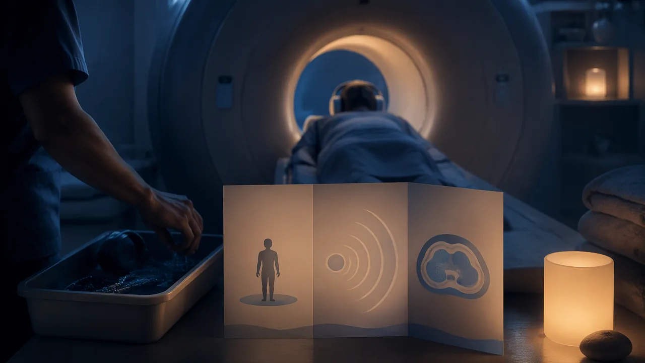
explain accurately how one diagnostic tool or imaging method works and what a patient experiences, and design a tri-fold brochure whose reading order serves the patient.
Instruction
A patient brochure is one of the most common pieces of media design in health care, and one of the most often done badly. Your brochure will explain one diagnostic tool or imaging method to a patient who is about to experience it. First the science, then the design.
Diagnostic tools. The stethoscope was invented by the French physician René Laennec in 1816; clinicians use it to listen to the heart, lungs and bowel. A heart murmur, for example, is an extra whooshing sound heard through the stethoscope, which may be harmless or may need further tests. The sphygmomanometer measures blood pressure with an inflatable cuff; the mercury cuff device was introduced by Scipione Riva-Rocci in 1896, and in 1905 Nikolai Korotkoff described the sounds heard through a stethoscope as the cuff is released, which give the systolic and diastolic readings. Many clinics now use automatic cuffs.
Imaging methods, as described by the NIH National Institute of Biomedical Imaging and Bioengineering. X-ray imaging, discovered by Wilhelm Röntgen in 1895, passes a small dose of ionizing radiation through the body; dense tissues like bone absorb more and appear white. Computed tomography (CT) takes X-ray measurements from many angles and uses a computer to build cross-sectional images; the first clinical CT scan was made in 1971 with the scanner developed by Godfrey Hounsfield. CT uses more radiation than a single X-ray, which clinicians weigh against the benefit. Magnetic resonance imaging (MRI) uses a strong magnet and radio waves, not ionizing radiation; because of the magnet, patients are screened for metal implants and objects. It is loud, and patients lie still in a tube, sometimes for a long time. Ultrasound sends high-frequency sound waves into the body and builds images from the echoes; it uses no ionizing radiation and is widely used in pregnancy.
A Missouri chapter. The Mallinckrodt Institute of Radiology at Washington University School of Medicine in St. Louis records that positron emission tomography (PET) was invented there in the 1970s by Michael Phelps and Michel Ter-Pogossian, and that in 1974 the institute acquired one of the first six CT scanners in the United States. As NIBIB explains, a PET scan uses a radioactive tracer; positrons from the tracer meet electrons in the body and produce pairs of gamma-ray photons, which the scanner’s detectors measure to build images of processes inside the body.
What the patient wants to know. Patients ask: what is it for, what will happen, will it hurt, how long will it take, is it safe, and when will I know the result? Answer those, in that kind of order, with facts from your sources. Do not give medical advice and do not promise results.
Tri-fold structure. A letter-size sheet folded in three has six panels. The front cover (outside right) carries the title and one image. The inside flap (inside left) is read first when the brochure opens: use it for “what is it and why”. The inside middle and inside right carry “what will happen”, step by step with small illustrations. The outside middle (the back) holds sources and contact information. Sketch a folded paper mock-up first and number the panels, because the reading order is not left to right on the flat sheet.
Design. Use a readable body size (at least 11 points in print), short paragraphs, headings written as the patient’s questions, and illustrations that show the patient’s experience, such as someone lying on the MRI table, rather than only the machine.
📖 Read for this lesson
The reading opens over the lesson; close it to come back to exactly where you were. The checks below draw on it.
Formative check
Work through these before moving on. They are not graded — they tell you whether the standard below has landed yet.
Which imaging methods use no ionizing radiation?
Korotkoff’s 1905 description of the sounds heard as a blood pressure cuff deflates is the basis of manual systolic and diastolic readings.
When a tri-fold brochure is opened, which panel does the reader usually see first?
List your six panels in reading order with the heading for each, written as a patient’s question. Name the source for the ‘Is it safe?’ panel.
How confident are you that you can explain a diagnostic tool or imaging method accurately and design a brochure with a patient-centered reading order?
Practice
Work these on paper or in your notebook, then open Check your answer. Aim for all of Fluency and Application; try at least one Challenge.
Printable version: this unit’s practice workbook (PDF)
Fluency
Build speed and accuracy with the core skill.
- Match each method to how it makes an image: X-ray, CT, MRI, ultrasound. Choices: echoes of high-frequency sound; a strong magnet and radio waves; X-ray measurements from many angles combined by a computer; a small dose of ionizing radiation through the body.
Check your answer
Answer: X-ray: a small dose of ionizing radiation through the body. CT: X-ray measurements from many angles combined by a computer into cross-sections. MRI: a strong magnet and radio waves. Ultrasound: echoes of high-frequency sound waves.From the NIBIB descriptions in the lesson. - Give the person and year for each: stethoscope; mercury cuff device; the sounds used for manual systolic and diastolic readings; discovery of X-rays; first clinical CT scan.
Check your answer
Answer: Stethoscope: René Laennec, 1816. Mercury cuff device: Scipione Riva-Rocci, 1896. Sounds: Nikolai Korotkoff, 1905. X-rays: Wilhelm Röntgen, 1895. First clinical CT scan: 1971, with the scanner developed by Godfrey Hounsfield.A timeline of these would run from 1816 to 1971. - List the six questions the lesson says patients ask.
Check your answer
Answer: What is it for? What will happen? Will it hurt? How long will it take? Is it safe? When will I know the result?Answer them in that kind of order, with facts from sources; do not give medical advice or promise results.
Application
Use the skill in context. Show your reasoning.
- Put the six panels of a tri-fold in reading order and give the job of each, following the lesson.
Check your answer
Answer: 1. Front cover (outside right): title and one image. 2. Inside flap (inside left): what is it and why. 3. Inside middle and 4. inside right: what will happen, step by step with small illustrations. 5. Outside middle (back): sources and contact information. The sixth panel is the remaining outside panel (the flap that folds in), used for a supporting topic such as ‘Is it safe?’.The lesson assigns five panel roles; the sixth is a design choice. Reading order is not left to right on the flat sheet, so make a folded mock-up and number the panels. - Rewrite these brochure headings as the patient’s questions: (a) ‘Procedure’; (b) ‘Safety information’; (c) ‘Duration’.
Check your answer
Answer: (a) ‘What will happen during my scan?’ (b) ‘Is it safe?’ (c) ‘How long will it take?’Headings written as the patient’s questions match what patients want to know. - A brochure about MRI says: ‘MRI uses a small dose of radiation, like an X-ray.’ Correct it using the lesson, and add one fact a patient should know before the scan.
Check your answer
Answer: Correction: MRI uses a strong magnet and radio waves, not ionizing radiation. Facts a patient should know: because of the magnet, patients are screened for metal implants and objects; the scan is loud, and the patient lies still in a tube, sometimes for a long time.Check every claim against the source; X-ray and CT use ionizing radiation, MRI and ultrasound do not. - A letter-size sheet is 11 inches wide. Folded into three equal panels, how wide is each panel to two decimal places? If body text is 11 points and 72 points make an inch, about how tall is one line of 11-point type in inches?
Check your answer
Answer: About 3.67 inches per panel; about 0.15 inch.11 / 3 = 3.667, about 3.67. 11 / 72 = 0.153, about 0.15. The lesson’s minimum body size for print is 11 points.
Review
Keep earlier skills sharp.
- Review 3.1: CT builds cross-sectional images. Which anatomical view type from Unit 3 matches a CT image, and how would you illustrate it on the brochure?
Check your answer
Answer: The cross-section, a view of the body as if sliced. Illustrate it with a simple sliced-body diagram, beside an image of the patient lying on the CT table to show the patient’s experience.The lesson asks for illustrations of the patient’s experience, not only the machine. - Review 4.3: Your brochure includes an example scan image. Why should it be an openly licensed or illustrative image rather than a scan of a real, identifiable patient?
Check your answer
Answer: A real image could carry information that identifies a person; use illustrative or public-domain images, and remove any attached patient information.Imaging data is stored with information about the patient attached.
Six Diagnostic Tools with Sources, a Worked Six-Panel MRI Brochure Plan and Print Measurements
SLS original brochure plan and measurements; tool facts from NIBIB pages (Magnetic Resonance Imaging; Computed Tomography; Ultrasound; X-rays fact sheets), NIH PubMed Central history articles on the stethoscope, Riva-Rocci and Korotkoff, the American Physical Society (Nov. 8, 1895) and NobelPrize.org/ACNR (Hounsfield, 1971); retrieved Sept. 26, 2026 · SLS original, paraphrasing public-domain NIH pages
Why this reading. The workshop asks you to explain one tool accurately and to name the source for the “Is it safe?” panel, but the lesson gives one sentence per tool. This reading gives checked facts and dates for six tools with their sources, a full worked six-panel plan for an MRI brochure, and the print measurements. The brochure plan is an SLS original example. Nothing here is medical advice, and a real brochure would be reviewed by a clinic.
Facts you may use, with sources
| Tool | What I can state, and its source | Where it came from |
|---|---|---|
| Stethoscope | Invented by René Laennec at the Necker Hospital in Paris; he made the first one from a tube on 13 September 1816. Clinicians listen through it to the heart, lungs and bowel. | History of the stethoscope articles in NIH PubMed Central (for example PMC1279298); the uses are from the lesson |
| Blood pressure cuff | Scipione Riva-Rocci introduced the upper-arm cuff in 1896, which measured systolic pressure by raising cuff pressure until the pulse could not be felt; in 1905 Nikolai Korotkoff added listening with a stethoscope as the cuff is released, so both systolic and diastolic pressure can be read. | PubMed and PMC history articles on Riva-Rocci and Korotkoff (for example PMC11520479, PubMed 16340219) |
| X-ray | X-rays pass through the body and are absorbed in different amounts by different tissues; bone, which contains calcium, absorbs more and appears whiter, while fat, muscle and air-filled lungs appear in shades of gray. Wilhelm Röntgen produced and detected X-rays on 8 November 1895. | NIBIB X-ray fact sheets; American Physical Society, “November 8, 1895” |
| CT scan | A narrow beam of X-rays is aimed at the patient and rotated around the body; a computer processes the signals into cross-sectional images (slices). The patient lies on a bed that slowly moves through the ring. CT uses X-rays, and all X-rays produce ionizing radiation. Some scans use an iodine-based contrast agent injected into a vein. The first CT scan of a patient was made on 1 October 1971 with Godfrey Hounsfield’s machine at Atkinson Morley Hospital. | NIBIB, Computed Tomography; ACNR / NobelPrize.org 1979 |
| MRI | A powerful magnet aligns protons in the body; radio-frequency pulses knock them out of alignment, and sensors detect the energy released as they realign, which tells tissues apart. MRI does not use the ionizing radiation of X-rays. The patient lies inside a large magnet and must stay still to avoid a blurred image. Scanners are loud (clicking and beeping, up to 120 decibels in certain scanners), so ear protection may be needed. People with certain implants, especially iron-containing ones such as pacemakers, should not enter an MRI machine, so screening comes first. | NIBIB, Magnetic Resonance Imaging |
| Ultrasound | A transducer sends high-frequency sound into the body and detects the echoes that bounce back from boundaries between tissues; the scanner uses the speed of sound and the time each echo returns to build an image. A gel is applied to the skin so no air pocket blocks the sound. Diagnostic ultrasound is generally regarded as safe and produces no ionizing radiation. It is used in pregnancy and for the heart, blood vessels, eyes, thyroid, breast, abdominal organs and more. | NIBIB, Ultrasound |
All dates and statements were checked on 26 September 2026 against the sources named; the National Institute of Biomedical Imaging and Bioengineering (NIBIB) pages are U.S. government works (public domain). I paraphrase them. If you use a different tool, find your own sources and record them the same way.
The six panels of a worked example: an MRI brochure
| Panel | Position | Heading (a patient’s question) | Content, from the sources above |
|---|---|---|---|
| 1 | Front cover, outside right | What is an MRI scan? | Title, one image of a person lying on the table with the ring behind, the sentence: a magnet and radio waves make pictures of the inside of your body |
| 2 | Inside flap (read first on opening) | Why am I having this scan? | Plain-language reason: a doctor asked for pictures; ends with “Ask your doctor why yours was ordered” (no medical advice) |
| 3 | Inside middle | What will happen? | Steps with small drawings: metal checklist, lie on the table, stay still, sounds, done |
| 4 | Inside right | Will it hurt? How loud is it? | Painless in itself; loud clicking and beeping, ear protection may be given; you must stay still |
| 5 | Outside left (folded in) | Is it safe? | No X-ray-type radiation; the strong magnet is why staff ask about implants such as pacemakers; source printed on the panel |
| 6 | Outside middle (back) | Where can I learn more? | Sources with dates, and a line for the clinic’s own phone number to fill in |
A note on panel order. The lesson calls the panel read first the inside flap, and puts the front cover at the outside right and the sources on the outside middle. The physical fold decides which panel meets the reader’s eye, so make the folded mock-up (below), number each panel in pencil, then check that a reader who opens the cover meets panel 2, in the order the lesson describes; if yours differs, renumber and reorder your content to match your mock-up.
Measurements for a letter tri-fold
Sheet: US Letter, 11 × 8.5 in, landscape, printed both sides. Each panel is about 3.67 in wide (11 ÷ 3); many templates make the panel that folds inward slightly narrower, about 3.6 in, so it does not buckle. Keep a 0.25 in safe margin inside each panel, leaving about 3.17 in for text. Body type at least 11 points (from the lesson), headings 16 to 20 points, phrased as questions. Aim for 50 to 80 words a panel and one illustration, so the text is short paragraphs (an SLS planning guide, not a standard). Folded mock-up: fold a letter sheet in thirds, unfold, number the six panels lightly, draw a box in each, then fold again and check the numbers appear in the order you planned when you open it.
Answering “is it safe?” without giving advice
State what the source says, name it, and hand medical questions back to the clinic. Worked panel 5 text (SLS original, from the NIBIB facts above): “An MRI does not use the X-rays of a CT scan or an X-ray. It uses a strong magnet, so the staff will ask whether you have any metal in your body, such as a pacemaker. Please tell them. Source: National Institute of Biomedical Imaging and Bioengineering, Magnetic Resonance Imaging. If you have a question about your own health, ask your doctor.” Notice it makes no promise about results and gives no advice for any one patient.
Sources: NIBIB, Magnetic Resonance Imaging (MRI) (nibib.nih.gov); NIBIB, Computed Tomography (CT) (nibib.nih.gov); NIBIB, Ultrasound (nibib.nih.gov); NIBIB, X-rays fact sheet (nibib.nih.gov); An Ear to the Chest: an Illustrated History of the Evolution of the Stethoscope (PMC1279298) (pmc.ncbi.nlm.nih.gov); From Concept to Cure: The Life and Legacy of Scipione Riva-Rocci (PMC11520479) (ncbi.nlm.nih.gov); A centenary of auscultatory blood pressure measurement: a tribute to Nikolai Korotkoff (pubmed.ncbi.nlm.nih.gov); American Physical Society, November 8, 1895: Roentgen’s Discovery of X-Rays (aps.org); NobelPrize.org, Physiology or Medicine 1979: Perspectives (nobelprize.org)