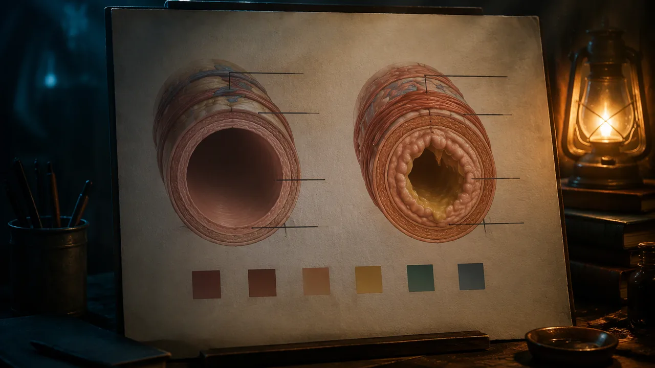
design an accurate, accessible illustrated panel that compares a healthy structure with one affected by a common condition, using labeling and color conventions deliberately.
Instruction
Your atlas panel is a piece of scientific illustration with an artist’s voice. This workshop covers the craft decisions that make such a panel accurate, readable and yours.
The comparison. The most powerful structure for explaining a condition is normal beside affected, drawn at the same scale and from the same view so that the only difference is the one that matters. Take asthma. The NIH National Heart, Lung, and Blood Institute explains that in asthma the airways become inflamed and swollen, the muscles around them tighten, and extra mucus can form, all of which narrow the path for air. A cross-section of a small airway shows this well: on the left, a round open airway with a thin wall; on the right, the same airway with a thicker, swollen wall, tightened muscle band and mucus reducing the open space. A viewer grasps the cause of wheezing and breathlessness without reading a paragraph.
Labels and leader lines. Put labels outside the drawing, aligned in a neat column, with thin straight leader lines that do not cross. Use everyday words first and the technical term second: “small airway (bronchiole)”. Carter’s direct labeling in Gray’s Anatomy is an alternative when parts are large enough.
Color. Choose a limited palette with a job for each color. Use value (light and dark) as well as hue, so the panel still works in grayscale. Avoid relying on red-green differences; about one in twelve men and a much smaller share of women have some form of red-green color-vision deficiency. Test by converting a photo of your panel to grayscale.
Type and caption. Choose one clear sans-serif typeface for labels and one for headings. Write a caption of 40-60 words in plain language: what the condition is, what changes in the body and how it is found. Put the most important sentence first.
Diagnostic note. Add a small inset showing how clinicians detect the condition. For asthma this includes listening to the lungs and a breathing test called spirometry, which measures how much air a person can breathe out and how fast. For high blood pressure it is measurement with a blood pressure cuff. Check the diagnostic methods for your condition on MedlinePlus or the relevant NIH institute page.
Accuracy check. Before you ink or finalize a digital file, set the draft aside for a day, then check every label against OpenStax and every claim about the condition against your federal source, ticking each one on a list. Avoid claims about cures, causes or treatments unless your source states them; this panel explains, it does not advise.
Tools. Pencil, fine liners and colored pencil on bristol board, or a free program: Inkscape for crisp vector diagrams, Krita for painted illustration. Scan or photograph hand work at 300 pixels per inch if possible.
📖 Read for this lesson
The reading opens over the lesson; close it to come back to exactly where you were. The checks below draw on it.
Formative check
Work through these before moving on. They are not graded — they tell you whether the standard below has landed yet.
Why should the normal and affected drawings be the same scale and view?
According to NHLBI, in asthma the airways can become inflamed and swollen, surrounding muscles can tighten and extra mucus can form.
Which label follows the plain-language convention for the panel?
Paste your 40-60 word caption. Then list each label on your diagram with the source you checked it against.
How confident are you that you can design an accurate, accessible comparison panel for a condition of one body system?
Practice
Work these on paper or in your notebook, then open Check your answer. Aim for all of Fluency and Application; try at least one Challenge.
Printable version: this unit’s practice workbook (PDF)
Fluency
Build speed and accuracy with the core skill.
- Define leader line, value and spirometry.
Check your answer
Answer: Leader line: a thin line connecting a label to the structure it names. Value: the lightness or darkness of a color, which carries contrast even in grayscale. Spirometry: a breathing test that measures how much air a person can breathe out and how fast.Spirometry is one of the diagnostic methods the lesson names for asthma. - What are the lesson’s rules for labels? Give three.
Check your answer
Answer: Put labels outside the drawing in a neat column; use thin straight leader lines that do not cross; use everyday words first and the technical term second, such as ‘small airway (bronchiole)’.Carter’s direct labeling is an alternative when parts are large enough. - How long should the panel’s caption be, and what three things should it say?
Check your answer
Answer: 40-60 words in plain language: what the condition is, what changes in the body, and how it is found, with the most important sentence first.The panel explains; it does not advise.
Application
Use the skill in context. Show your reasoning.
- Describe the normal-beside-affected comparison for asthma the lesson suggests: what view, and what three differences appear in the affected airway?
Check your answer
Answer: A cross-section of a small airway, same scale and view on both sides. The affected airway shows a thicker, swollen wall, a tightened muscle band, and mucus reducing the open space.These match NHLBI’s description: inflamed and swollen airways, tightened muscles and extra mucus, which narrow the path for air. - About one in twelve men has some form of red-green color-vision deficiency. In an invented audience of 600 men, about how many is that? What test does the lesson recommend for your panel?
Check your answer
Answer: About 50 men. Convert a photo of the panel to grayscale and check it still reads.600 / 12 = 50. Using value as well as hue keeps the panel readable. - You scan a hand-drawn panel that is 8 inches by 10 inches at 300 pixels per inch. What are the pixel dimensions, and how many pixels in total?
Check your answer
Answer: 2,400 by 3,000 pixels; 7,200,000 pixels (7.2 megapixels).8 x 300 = 2,400; 10 x 300 = 3,000; 2,400 x 3,000 = 7,200,000. - Write a 40-60 word plain-language caption for an invented panel on high blood pressure. Use only facts from the unit: it often has no symptoms and is found by measurement with a blood pressure cuff.
Check your answer
Answer: Model (47 words): ‘High blood pressure is a condition of the cardiovascular system, the heart and blood vessels. It often causes no symptoms at all, so a person can feel fine and not know it is there. That is why it is found by measurement, using a blood pressure cuff.’Count the words (40-60), put the most important point first (often no symptoms, found by measurement), and add no claims about causes, cures or treatments. Every fact in the model comes from the unit: the system, no symptoms, measurement with a cuff.
Challenge
Stretch problems. Expect to think before you write.
- A draft caption says: ‘Asthma is caused by stress and can be cured by breathing exercises.’ Using the accuracy check, explain what is wrong and rewrite it from the unit’s facts only.
Check your answer
Answer: The lesson’s source does not state these causes or a cure, and the panel must not make claims about causes, cures or treatments unless the source states them. Rewrite: ‘In asthma, the small airways become swollen, the muscles around them tighten and extra mucus can form, narrowing the path for air. Clinicians find it by listening to the lungs and with a breathing test called spirometry.’Check every claim against the federal source; the panel explains, it does not advise.
Review
Keep earlier skills sharp.
- Review 3.2: Which examination technique is listening with a stethoscope, and which body sounds does the lesson say it is used for?
Check your answer
Answer: Auscultation; the heart, lungs or bowel.For asthma, the panel’s diagnostic inset includes listening to the lungs.
Technique Sheet: A Normal-Versus-Affected Asthma Panel with Measurements, Labels, Color Test and Caption
SLS original technique sheet; asthma facts paraphrased from NHLBI pages (Asthma; Asthma Causes) and color-vision prevalence from the National Eye Institute, retrieved Sept. 26, 2026; contrast ratios computed with the WCAG 2.1 formula · SLS original, paraphrasing public-domain NIH pages
Why this reading. The workshop asks you to draw a normal-versus-affected panel, label it, choose colors, and write a 40–60 word caption, and it cites the NIH institute’s account of asthma without giving it. This is the technique sheet: the NHLBI wording, a page layout with measurements, a schematic you can draw with a ruler and compass, a label list with sources, a color test with computed values, and a caption that fits the count. The layout, drawing proportions and caption are SLS originals; the proportions are schematic, not measurements of a real airway.
What the source says
The National Heart, Lung, and Blood Institute (NHLBI) says asthma is a chronic (long-term) condition that affects the airways in the lungs; that during an episode the airways swell, narrow and can create more mucus; that the muscles around the airways may also tighten; and that over time the airway walls can become thicker. (NHLBI asthma pages, retrieved 26 September 2026; public domain.) Those three changes are exactly what your affected drawing has to show, and nothing else in the drawing should change.
Page layout, in inches
US Letter, landscape, 11 × 8.5, margins 0.5. Title band across the top, 1 in tall. Below it, left to right across the 10 in live width: left label column 1.5; normal drawing frame 3.5; gutter 0.3; affected drawing frame 3.5; right label column 1.2 (1.5 + 3.5 + 0.3 + 3.5 + 1.2 = 10.0). Frames are 4.2 in tall. A caption box 10 in wide and 1.3 in tall runs along the bottom. Type: heading 28 pt, labels 12 pt, caption 11 pt, source line 9 pt.
The same-scale schematic (SLS original proportions)
Draw both airways as cross-sections on the same scale, one in each frame, each centered in a 3.5 × 4.2 in frame. Use a compass and a scale of 1 mm on the page per unit below.
| Part | Normal airway | Affected airway (same view, same scale) |
|---|---|---|
| Outer edge of the airway wall (circle diameter) | 60 mm | 60 mm |
| Wall thickness, lining | 4 mm | 12 mm (swollen) |
| Muscle band (ring outside the lining) | 3 mm, loose | 3 mm, drawn tighter with crinkled edge |
| Open space for air (circle diameter left inside) | 52 mm | 36 mm, plus mucus in the lower third |
| Area of open circle | about 2,124 mm² (π × 26²) | about 1,018 mm² (π × 18²), less the mucus |
The open space of the affected drawing is about 48% of the normal one before the mucus is subtracted (1,018 ÷ 2,124). These numbers are a drawing rule so the difference is visible, not a claim about real airways; the caption says “drawings are simplified.” The same scale and view keep the only visible difference the one that matters.
Labels and leader lines
Put labels outside the drawing in an aligned column, with thin straight leader lines that do not cross; everyday words first, technical term second. Worked list, each with its source:
| Label on the panel | Where it points | Source I checked |
|---|---|---|
| Small airway (bronchiole) | The ring in both drawings | The lesson’s term; confirm “bronchiole” on a federal page before you print it |
| Airway lining, swollen | The thick inner ring on the right | NHLBI: the airways swell |
| Muscle around the airway, tight | The outer band on the right | NHLBI: the muscles around the airways may tighten |
| Extra mucus | The blob in the lower third on the right | NHLBI: can create more mucus |
| Open space for air | The empty circle in both | Drawing; unlabeled claim, no source needed |
Color, tested with numbers
The National Eye Institute says about 1 in 12 men have color vision deficiency, and the most common type makes it hard to tell red from green. So do not use red versus green as your only difference. Test by converting a photo to greyscale, or with this calculation for a pair of colors: the WCAG contrast ratio. Two examples I computed with the WCAG formula: red #B33A3A and green #3A7D44 have a contrast of only 1.17 to 1 (their gray levels are about 94 and 98 out of 255, so they look the same in greyscale); navy #1F4E79 and amber #F2B134 have a contrast of 4.58 to 1 (gray levels about 69 and 182). Use navy for the lining and amber for the mucus, or add a pattern (stripes for mucus).
The caption
Write 40 to 60 words in plain language: what the condition is, what changes in the body, how it is found. Put the most important sentence first. Worked caption (SLS original, 53 words): “Asthma is a long-term condition of the airways in the lungs. In a flare-up the airway wall swells, the muscles around it tighten, and extra mucus can form. The open space for air gets smaller, so breathing is harder. Doctors can listen to the lungs with a stethoscope. Drawings are simplified. Source: NHLBI.”
Check yours: count words, then test each sentence for its source. In this example the first three sentences rest on NHLBI, the fourth on the lesson’s description of the stethoscope as a tool clinicians use to listen to the lungs, and the last two are honesty statements.
Sources: NHLBI, Asthma (nhlbi.nih.gov); NHLBI, Asthma Causes and Risk Factors (nhlbi.nih.gov); National Eye Institute, Color Blindness (nei.nih.gov)