1.05 Proteins at Work: Enzymes and Temperature
explain how an enzyme’s shape determines its function, measure how temperature affects catalase activity, and use the data to model why the body holds its temperature in a narrow range.
- I can explain how genes, which are regions of DNA, determine the structure of proteins that carry out essential functions in specialized cells.
- I can use reasoning to connect evidence and scientific principles to explain how genes in DNA code for proteins in all cells.
- I can describe how specialized cells in tissues rely on proteins to carry out their functions, and how proper protein function is necessary for cells to work correctly.
- I can explain how changes in gene sequences can affect protein function and, in turn, the function of body tissues.
Phase 3: Make Sense
Warm-Up ~5 min
Do Now. A raw egg white is clear and runny. Cooked, it is white and firm, and it stays that way when it cools. What do you think changed in its proteins?
You boil a yeast suspension, cool it to 37 °C, and add hydrogen peroxide. What will happen?
Instruction ~10 min
Proteins do the work. Almost every job inside a cell is done by a protein: enzymes speed reactions, channel proteins let ions cross membranes, hemoglobin carries oxygen, and keratin toughens skin. A protein is a chain of amino acids, and the sequence of amino acids, set by a gene (a region of DNA), determines how the chain folds into a particular three-dimensional shape. Shape determines function. You will trace how DNA sets the amino acid sequence in Unit 3.
Specialized cells, specialized proteins. Every body cell carries the same genes, but each type uses different ones. Sweat-gland cells make channel and pump proteins that move salt and water; red blood cell precursors make hemoglobin; muscle cells make the motor proteins that contract. When a gene’s sequence changes, its protein can change too. In sickle cell disease, one change in the hemoglobin gene swaps a single amino acid; the altered hemoglobin clumps when oxygen is low, red blood cells bend into a sickle shape, and blood flow through small vessels suffers. One gene, one protein, a whole tissue affected.
How enzymes work. An enzyme is a protein catalyst: it speeds a reaction without being used up. The molecule it acts on, the substrate, fits into a pocket called the active site, where the enzyme’s shape stresses the substrate’s bonds and lowers the energy needed for the reaction to start. Because each active site has a specific shape, each enzyme acts on one substrate or a few similar ones.
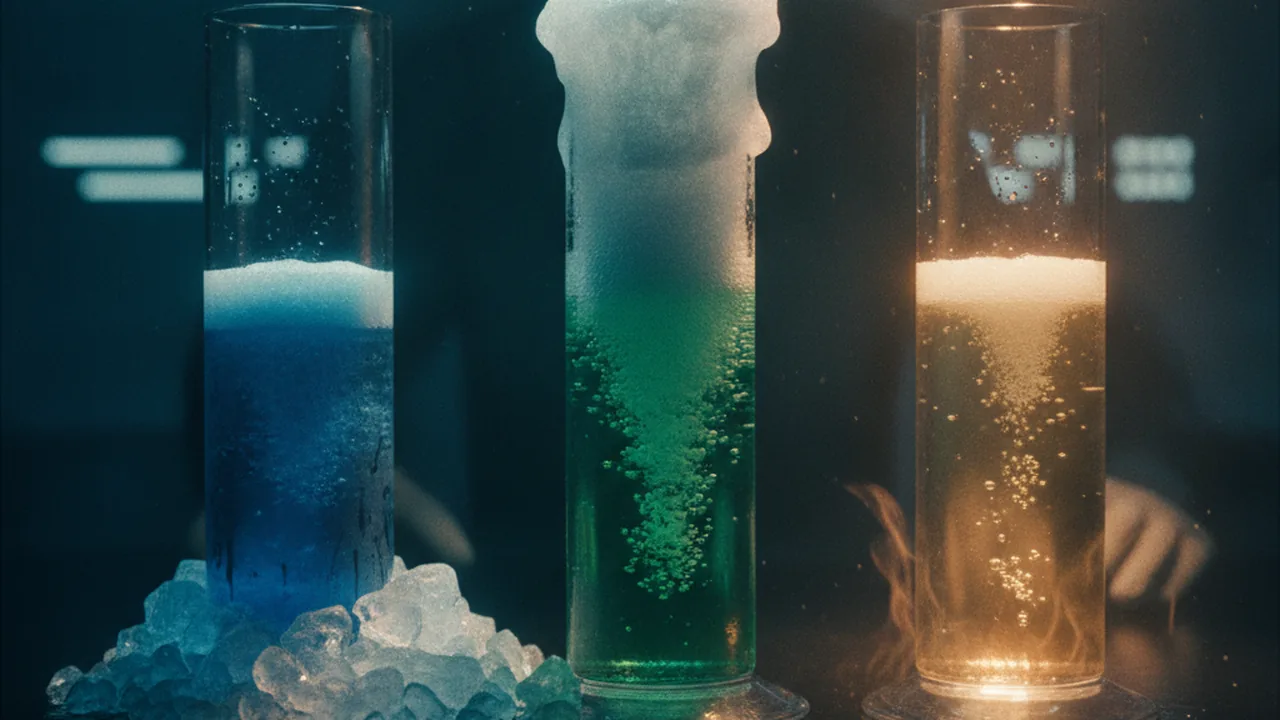
Catalase. Cells produce hydrogen peroxide (H2O2) as a by-product of normal chemistry, and it damages cell parts. The enzyme catalase breaks it down: 2H2O2 → 2H2O + O2. Yeast cells are full of catalase, so when you mix yeast with 3% hydrogen peroxide, oxygen bubbles form foam. The height of the foam in a set time is a measure of enzyme activity.
Temperature and shape. Warming speeds molecules, so enzyme and substrate collide more often and the reaction goes faster, up to a point. Above an optimum, the weak bonds holding the protein in shape break, the active site loses its shape, and the enzyme is denatured. Denaturing is often permanent: a cooked egg white does not become clear again when it cools.
Worked example 1: rates from a graph (sample data). Yeast catalase, foam height after 60 s: 4 °C, 12 mm; 22 °C, 25 mm; 37 °C, 38 mm; 50 °C, 20 mm; 80 °C, 3 mm. Between 22 and 37 °C foam rose 13 mm over 15 °C, about 0.87 mm per °C; between 37 and 50 °C it fell 18 mm over 13 °C, about 1.4 mm per °C. The fall after the optimum is steeper than the rise before it, because denaturing destroys enzymes rather than just slowing them.
Worked example 2: reading a graph honestly. Five temperatures cannot locate an optimum exactly. The data show it lies somewhere between 22 and 50 °C, most likely near 37 °C. To narrow it, add trials at 30, 40 and 45 °C and repeat each at least three times. Judging whether your data are precise and complete enough is part of every investigation.
Building the model. Draw the enzyme with its active site at three temperatures: cold (correct shape, slow collisions), near the optimum (correct shape, fast collisions) and hot (unfolded, the substrate no longer fits). Then link the model to the phenomenon. Human enzymes work best near 37 °C. At a core temperature of 40 °C and above, some proteins begin to lose their shape, cells stop working properly, and the brain, which runs the feedback loop, is among the first organs affected. The negative feedback you measured in 1.04 protects the shapes of the proteins that keep you alive.
Lab procedure: catalase activity in yeast at different temperatures.
- Pour 5 mL of yeast suspension (1 packet of dry yeast in 100 mL warm water, rested 10 minutes) into each of five tubes and place one in each water bath (about 4, 22, 37, 50 and 80 °C) for five minutes. Record the actual temperatures.
- Measure 5 mL of 3% hydrogen peroxide into each of five matching tubes and warm or cool them in the same baths.
- Pour the peroxide into the yeast for one temperature, start the stopwatch, and after 60 seconds measure the foam height in millimeters above the liquid.
- Repeat for each temperature, keeping volumes, timing and tube size the same.
- Repeat the whole set at least twice (class groups can share the work) and calculate the mean foam height for each temperature.
| Temperature (°C) | Trial 1 foam (mm) | Trial 2 (mm) | Trial 3 (mm) | Mean (mm) |
|---|---|---|---|---|
| about 4 | ||||
| about 22 | ||||
| about 37 | ||||
| about 50 | ||||
| about 80 |
Reading: OpenStax Biology 2e, section 6.5, Enzymes (CC BY 4.0).
Guided Exercises ~15 min
Graph the class means first, then work through these.
Independent Practice ~10 min
Finish the analysis in your notebook.
Name the independent, dependent and three controlled variables. Evaluate your data: how precise were repeated trials, what is one limitation, and what would you change to make the optimum more certain?
Explain the chain from gene to function: how does the DNA sequence of the catalase gene end up deciding whether yeast can break down peroxide at 37 °C?
Closure ~5 min
Proteins explain why the body guards 37 °C. Next, we pull the whole unit’s science into one model.
Exit ticket: use your graph to explain why a core temperature of 40 °C or more is dangerous to a person.
How confident are you that you can explain how an enzyme's shape determines its function and why high temperature destroys that function.
Teacher notes
Materials: dry baker’s yeast, 3% hydrogen peroxide (drugstore strength), five clear tubes or cups per group, rulers, 10 mL syringes or spoons, ice bath, room-temperature water, warm tap water (~37 °C), hot tap water (~50 °C), an electric kettle for ~80 °C water (teacher only), thermometers, stopwatches, goggles.
Timing: this lab spans two 50-minute periods: data on day 1, graph, model and analysis on day 2. Sample data are available for groups whose trials fail.
Listen for: “heat kills the enzyme” (enzymes are not alive; they denature) and “cold denatures enzymes too” (cold slows them; shape usually survives).
Support / stretch: give axis labels and a scale. Stretch: explain why a fever of 38.5 °C may help fight infection while 41 °C is dangerous.