1.03 Cells Up Close: Onion, Elodea and Salt Water
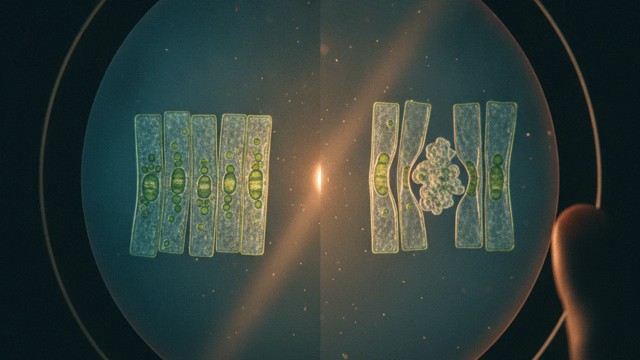
prepare wet-mount slides, identify cell structures, estimate cell size from the field of view, and explain what happens to plant cells in salt water in terms of osmosis.
- I can identify and describe evidence that all cells contain DNA.
- I can describe how DNA contains genes, and how gene sequences provide instructions for making proteins.
- I can explain how groups of specialized cells use proteins to perform essential functions for the organism.
- I can develop a model that identifies and describes the main organ systems, organs, and tissues in a multicellular organism.
- I can use my model to describe key processes, such as how fluids are transported or how movement occurs, within these body systems.
Phase 2: Investigate
Warm-Up ~5 min
Do Now. Lettuce left in salad dressing goes limp in an hour. Write one guess about what is happening to its cells.
You put a leaf cell into very salty water. What happens to the water inside it?
Instruction ~10 min
Why start with cells? Every function you studied in the launch, from sweating to pumping blood, is carried out by cells. Cell structures are matched to their jobs, and one structure matters for every cell: the plasma membrane, which controls what enters and leaves.
Plant and animal cells. All eukaryotic cells have a plasma membrane, cytoplasm, a nucleus holding DNA, ribosomes that build proteins, and mitochondria that release energy from food. Bacteria have no nucleus, but their DNA sits in the cytoplasm, so DNA is found in every kind of living cell. (Mature mammal red blood cells lose their nucleus as they develop; they cannot make new proteins and live only about four months.) Plant cells add three structures you will see today: a rigid cell wall of cellulose outside the membrane, a large central vacuole filled with water, and, in green tissue, chloroplasts. Onion bulb cells have no chloroplasts because the bulb grows underground; the Elodea leaf does. The iodine stain makes each onion nucleus show up as a dark dot: visible evidence of where the cell’s DNA is kept.
Using the microscope. Total magnification is the eyepiece magnification times the objective magnification: a 10x eyepiece with a 40x objective gives 400x. The field of view (FOV), the diameter of the circle you see, shrinks as magnification rises, in inverse proportion.
Worked example 1: cell size from the FOV. If the FOV is 4.5 mm at 40x, then at 400x (ten times the magnification) it is 4.5 ÷ 10 = 0.45 mm, or 450 µm. If 1.5 onion cells span the 400x field end to end, each is about 450 ÷ 1.5 = 300 µm long. Always measure the low-power FOV with a clear ruler first, because microscopes differ.
Worked example 2: why are cells small? A cell exchanges materials through its surface, but its needs grow with its volume. For a cube of side s, surface area is 6s2 and volume is s3, so the ratio is 6/s. A 1-unit cube has a ratio of 6; a 2-unit cube has 3; a 3-unit cube has only 2. As a cell grows, each unit of volume has less membrane to serve it. That is one reason large organisms are made of many small cells rather than a few huge ones.
Osmosis. The membrane lets water cross freely but blocks most dissolved salt. Water moves by osmosis from where it is more concentrated (fewer dissolved particles) to where it is less concentrated (more dissolved particles). Put a plant cell in salty water and water leaves the vacuole; the membrane pulls away from the rigid wall. This is plasmolysis. In fresh water, water enters, the vacuole swells and pushes against the wall, making the cell firm (turgid). Animal cells have no wall, so in very dilute water they can swell and burst, which is one reason the body tightly regulates the salt and water in blood.
Connecting to the phenomenon. A worker who sweats heavily loses both water and salt. Drinking plain water replaces the water but not the salt; drinking nothing concentrates the body’s fluids. Either way, the concentration outside cells changes, and cells gain or lose water by osmosis. Homeostasis includes keeping that concentration steady.
Lab procedure: onion and Elodea cells, and what salt water does to them. Question: what structures can we see in plant cells, how big are they, and how does salt water change them?
- Measure the field of view at 40x by placing a clear ruler on the stage; record the diameter in millimeters.
- Peel the thin inner skin from an onion layer with forceps, lay it flat in a drop of water on a slide, add one drop of iodine at the edge, and lower a cover slip at an angle to avoid bubbles.
- Focus at 40x, then 100x, then 400x. Draw three or four cells at 400x and label the cell wall, nucleus and cytoplasm. Count how many cells fit end to end across the 400x field.
- Make a wet mount of one Elodea leaf in tap water. Draw it at 400x, labeling chloroplasts and the cell wall. Watch for chloroplasts moving in the cytoplasm.
- Place a drop of salt solution (1 teaspoon of table salt in 100 mL of water) at one edge of the Elodea cover slip and touch a paper towel to the opposite edge to draw it under. Observe for five minutes and draw the same cells again.
- Replace the salt solution with tap water the same way and observe for five more minutes. Record whether the change reverses.
| Measurement | Value |
|---|---|
| FOV at 40x (mm) | |
| FOV at 400x (mm, calculated) | |
| Onion cells across 400x field | |
| Estimated onion cell length (µm) | |
| Elodea in tap water: description | |
| Elodea in salt water after 5 min: description | |
| Elodea back in tap water: description |
No microscope? Use the illustrative 400x micrograph plate your teacher provides (a 450 µm field): one onion field and three Elodea fields before, during and after salt water. It earns full credit.
Reading: OpenStax Biology 2e, chapters 4, Cell Structure, and 5, Structure and Function of Plasma Membranes (CC BY 4.0).
Guided Exercises ~15 min
Use your drawings and data table.
Independent Practice ~10 min
Finish your lab analysis.
Show your FOV calculation and your estimate of onion cell length. Compare with two classmates' estimates, suggest why they differ, and name two sources of error and how you would reduce them.
A worker drinks only plain water after hours of heavy sweating. Predict which way water moves across the membranes of the worker's cells, and explain why.
Closure ~5 min
Cells are where homeostasis happens. Next, we measure a whole-body feedback loop.
Exit ticket: give one piece of evidence from today that cells contain DNA, and one reason cells stay small.
How confident are you that you can prepare a slide, estimate cell size from the field of view, and explain plasmolysis with osmosis.
Teacher notes
Materials: compound microscopes (40x, 100x, 400x), slides and cover slips, one onion, Elodea from an aquarium supplier, Lugol’s iodine or diluted food coloring, salt solution (1 tsp in 100 mL), paper towel, forceps, clear rulers, goggles.
Timing: this lab spans two 50-minute periods: slides and drawings on day 1, plasmolysis and analysis on day 2.
Listen for: “salt sucks the water out” (water moves by osmosis; salt does not pull); “the cell wall shrank” (the membrane pulls away from a rigid wall). PhET link: confirm the Membrane Transport sim loads on school devices before class; the printed diagram is the backup.
Support / stretch: provide a labeled reference drawing and a calculation frame. Stretch: calculate SA:V for a 10 µm and a 100 µm cube and explain what it means for a cell’s supply of oxygen.