How does the information in a gene get precisely converted into a protein, and how can even a tiny change in that genetic information lead to a different protein with altered function?
transcribe a DNA template into mRNA, translate the mRNA into amino acids with the genetic code, and explain how a change in one base can change a protein’s structure and function, with harmful, beneficial or neutral effects.
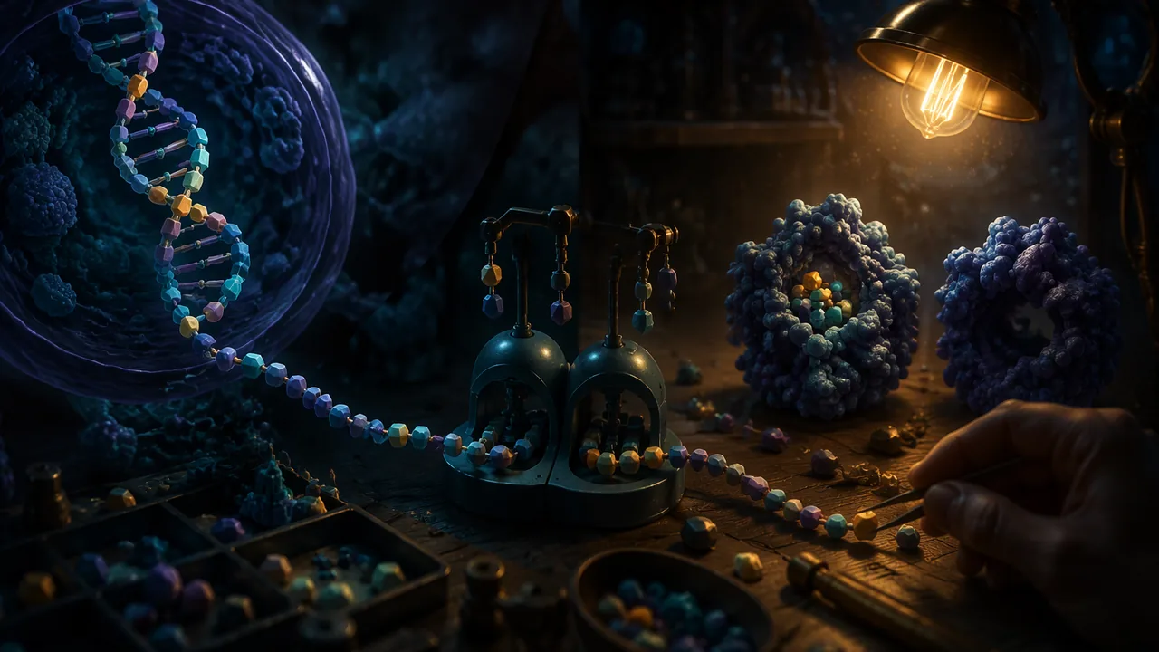
The central idea. DNA stays in the nucleus, but proteins are built on ribosomes in the cytoplasm. The cell copies a gene’s information into a messenger molecule and carries it out. The flow of information is DNA → RNA → protein. Proteins then do the work: enzymes such as catalase, structural proteins such as keratin, motor proteins such as myosin, and signals such as insulin. A protein’s function depends on its shape, and its shape depends on the order of its amino acids.
Genetic information flows from DNA to RNA to protein, where the sequence of amino acids determines a protein’s specific shape and function.
📖 Read for this lesson
The reading opens over the lesson; close it to come back to exactly where you were. The checks below draw on it.
Why is it important for DNA to stay protected in the nucleus while proteins are built elsewhere?
The DNA template TAC GGA CTT is transcribed into the mRNA .
Transcription
All known life forms on Earth, from bacteria to plants and animals, use essentially the same genetic code to translate mRNA into proteins.
An enzyme called RNA polymerase unzips a short section of DNA and builds a strand of messenger RNA (mRNA) that is complementary to one DNA strand, the template strand. RNA uses uracil (U) in place of thymine, so the pairing rules are: template A → mRNA U, T → A, G → C and C → G. Worked example: the template TAC AAA CCG TTT ATT is transcribed as AUG UUU GGC AAA UAA.
The mRNA codon GAG changes to GUG. Using the genetic code, what happens?
Translation
At the ribosome, the mRNA is read three bases at a time. Each triplet is a codon. Transfer RNA molecules carry amino acids and match their anticodons to the codons, and the ribosome links the amino acids into a chain. Why three bases? There are 20 amino acids. Codons of two bases would give only 42 = 16 combinations, not enough; three bases give 43 = 64, enough for all 20 plus start and stop signals. Using the standard genetic code table (OpenStax Biology 2e, section 15.1), our mRNA AUG UUU GGC AAA UAA reads: AUG = methionine (the start codon), UUU = phenylalanine, GGC = glycine, AAA = lysine, UAA = stop. The protein fragment is Met-Phe-Gly-Lys. Because several codons can code for the same amino acid (GAA and GAG both code for glutamic acid), the code is described as redundant.
A sequence of three mRNA bases that codes for one amino acid or a start or stop signal.
DNA leaves the nucleus to go directly to the ribosomes and build proteins.
DNA safely stays in the nucleus, acting as a protected blueprint. Messenger RNA (mRNA) carries a copy of the gene’s instructions to the ribosomes in the cytoplasm, where proteins are assembled.
Simulation & tools
Build a protein from a gene
Open Gene Expression Essentials and choose the Expression screen. (1) Drag the positive transcription factor and RNA polymerase onto the gene and record what is produced. (2) Place a ribosome on the mRNA and watch the protein grow; record how many proteins you make in one minute. (3) Remove the transcription factor and repeat: what changes, and why? Write two sentences explaining how a cell can carry a gene without making its protein. No internet? Use the transcription-factor paragraph in this lesson’s reading and answer the same questions.
Open in a new tab ↗ · PhET Interactive Simulations, University of Colorado Boulder · CC BY 4.0
When one base changes
A point mutation changes a single base. Its effect depends on where it lands:
- A silent mutation changes a codon to another codon for the same amino acid: GAA → GAG still gives glutamic acid.
- A missense mutation changes the amino acid. In sickle cell disease, the sixth codon of the β-globin gene changes from GAG to GUG, so glutamic acid becomes valine. That one change makes hemoglobin molecules stick together when oxygen is low, which bends red blood cells into a sickle shape.
- A nonsense mutation creates a stop codon early. AUG GCU UGG AAA UAG (Met-Ala-Trp-Lys) becomes AUG GCU UGA … if the G in UGG changes to A, and the chain stops after two amino acids.
- An insertion or deletion of one base shifts the reading frame, changing every codon after it.
A change in a single base of DNA; it may be silent, missense or nonsense.
Missouri standard: 9-12.LS3.B.2
A change in one base of a gene always harms the organism, because it always changes the protein.
Harmful, beneficial or neutral?
The effect on the organism depends on what the change does to the protein. MedlinePlus Genetics notes that most gene variants have no effect on health: a silent change, or a change that does not alter how the protein works, is neutral. Some are harmful: two copies of the GAG → GUG variant cause sickle cell disease. A very small share are beneficial, making a protein that helps the organism in its environment. The same hemoglobin S variant shows that one mutation can be both: according to the National Heart, Lung, and Blood Institute, having the hemoglobin S gene protects against serious malaria, which is why it is more common where malaria is common. Whether a mutation is harmful, beneficial or neutral depends on the protein change and on the environment.
Back to the lizard
A regrowing tail needs collagen and cartilage proteins in its new support tube, contractile proteins in its muscle and keratin in its scales. The cells all carry the same genes; each cell type transcribes a different set of them. The model you build today connects the level of the gene to the level of the tissue.
Key terms
Copying a gene’s DNA sequence into a complementary strand of messenger RNA.
Reading mRNA codons at the ribosome to assemble a chain of amino acids.
A mutation that inserts or deletes bases (not in a multiple of three), shifting how every later codon is read.
Lab: Paper and bead model of protein synthesis
Question. How does a sequence of DNA bases determine a sequence of amino acids, and what happens to the protein when one base changes?
Materials
- The four DNA template strands in this lesson’s reading, copied out by hand or printed, and blank strips for the mRNA
- Codon table card and amino-acid cards, or pop beads in 20 colors with a color key
- Scissors, tape and a pipe cleaner or a length of string
- Use scissors carefully and keep small beads off the floor to avoid slips.
- No chemicals are used; keep small beads away from young children and pets.
Procedure
- Place the DNA template strip on one spot on your table or desk labeled ‘nucleus’. Write the complementary mRNA bases on a blank strip, using U in place of T.
- Carry the mRNA strip to a second spot labeled ‘ribosome’. Mark off codons in groups of three from the start codon AUG.
- Use the codon table to pick the matching amino-acid card or bead for each codon, and thread the beads on the pipe cleaner in order until you reach a stop codon.
- Now mutate your gene. Choose one. All three earn full credit, and all three are set out for every gene in the table ‘Four gene strips and three mutations’ in this lesson’s reading: Option A changes one base of the template, Option B deletes one base, Option C changes a different base. Write the mutated template out in full, then repeat steps 1-3 with it.
- Compare the two chains and classify the mutation as silent, missense, nonsense or frameshift.
Data
| DNA template | mRNA | Amino-acid sequence | |
|---|---|---|---|
| Original gene | |||
| Mutated gene | |||
| Type of mutation | |||
Analysis
- Explain why a single-base deletion usually changes a protein more than a single-base substitution does.
- Use the sickle cell example to explain how a change in one amino acid can change the function of a protein and the shape of a cell.
- Decide whether the mutation you modeled would most likely be harmful, beneficial or neutral to the organism, and explain why, using the effect on the protein. Then explain how the hemoglobin S variant can be harmful in one situation and beneficial in another.
- Identify one way your paper model is unlike real transcription and translation, and explain why the difference matters or does not.
If you cannot do this lab in person: With no beads or cards, do the whole model on paper from the gene strips and codon table in this lesson’s reading, then check your chains with the PhET Gene Expression Essentials simulation.
Practice
Work these on paper or in your notebook, then open Check your answer. Aim for all of Fluency and Application; try at least one Challenge.
Printable version: this unit’s practice workbook (PDF)
Fluency
Build speed and accuracy with the core skill.
- Transcribe the DNA template TAC AAA CCG TTT ATT into mRNA.
Check your answer
Answer: AUG UUU GGC AAA UAAT→A, A→U, C→G, G→C. - Translate the mRNA AUG UUU GGC AAA UAA using the genetic code.
Check your answer
Answer: Met-Phe-Gly-Lys (then stop)AUG Met, UUU Phe, GGC Gly, AAA Lys, UAA stop. - Transcribe and translate the template TAC CTT AAA CCG ACT.
Check your answer
Answer: mRNA AUG GAA UUU GGC UGA; Met-Glu-Phe-Gly (then stop)GAA = Glu; UGA = stop. - How many different codons are possible with four bases taken three at a time?
Check your answer
Answer: 6443 = 64.
Application
Use the skill in context. Show your reasoning.
- A protein has 300 amino acids. How many mRNA bases code for it, including the stop codon?
Check your answer
Answer: 903300 × 3 = 900, plus 3 for the stop codon. - Classify each change to the mRNA codon GAA (Glu): (a) GAG; (b) GUA; (c) UAA.
Check your answer
Answer: (a) silent (still Glu); (b) missense (Val); (c) nonsense (stop)Use the genetic code: GAG = Glu, GUA = Val, UAA = stop. - Explain how the change from GAG to GUG in the β-globin gene changes the function of hemoglobin.
Check your answer
Answer: Glutamic acid (charged) is replaced by valine (nonpolar). The changed hemoglobin sticks together when oxygen is low, forming fibers that bend red blood cells into a sickle shape that can block small vessels.One amino acid changes the protein’s surface and therefore how molecules interact. - Label each change as most likely neutral, harmful or beneficial, and give the reason: (a) GAA → GAG in a gene for a muscle protein; (b) two copies of the GAG → GUG β-globin variant; (c) one copy of the same variant in a region where malaria is common.
Check your answer
Answer: (a) neutral: GAG still codes for Glu, so the protein is unchanged; (b) harmful: hemoglobin S causes sickle cell disease; (c) beneficial there: having the hemoglobin S gene protects against serious malaria.The effect depends on whether the protein changes and on the environment; the same variant can be harmful in one setting and beneficial in another.
Challenge
Stretch problems. Expect to think before you write.
- The mRNA AUG UUU GGC AAA UAA loses its seventh base (the first G of GGC). Write the new reading frame and the resulting amino acids, and explain why this mutation is more serious than a substitution.
Check your answer
Answer: AUG UUU GCA AAU AA…: Met-Phe-Ala-Asn, and the original stop codon is lost from the frame. Every codon after the deletion changes (a frameshift), whereas a substitution changes at most one amino acid.Remove the base and regroup into triplets: AUG UUU GCA AAU AA; GCA = Ala, AAU = Asn.
Review
Keep earlier skills sharp.
- Why must a cell copy its DNA before mitosis?
Check your answer
Answer: So that each daughter cell receives a complete set of chromosomes: each chromosome is copied into two sister chromatids that are separated in anaphase.Replication in S phase comes before division.
The ability of a lizard such as Missouri’s common five-lined skink to regrow a lost tail relies on cells accurately transcribing and translating specific genes to produce the precise collagen, cartilage, and muscle proteins needed for repair.
How does the redundancy of the genetic code offer some protection against the harmful effects of certain point mutations?
How confident are you that you can transcribe and translate a gene and explain how one base change can change a protein, and whether that is harmful, beneficial or neutral?
The Shift
- Genes are copied from DNA to mRNA in transcription.
- mRNA codons are read to build proteins in translation.
- Mutations can alter protein structure and function, with harmful, beneficial or neutral effects.
From Gene to Protein, and What One Base Can Do
SLS reading for RLP Biology A, drawing on MedlinePlus Genetics pages on protein synthesis, gene variants and the HBB gene, the National Heart, Lung, and Blood Institute on sickle cell disease and malaria, and the NHGRI Talking Glossary; retrieved November 5, 2026 · SLS original
Why a messenger is needed
DNA stays inside the nucleus, protected. Proteins are built on ribosomes out in the cytoplasm. So the cell copies the information in one gene into a portable molecule, messenger RNA (mRNA), and sends the copy out. The flow of information is DNA → RNA → protein. The protein then does the work: an enzyme such as catalase, a structural protein such as keratin or collagen, a motor protein such as myosin, or a signal such as insulin.
Step 1: Transcription (in the nucleus)
The enzyme RNA polymerase unzips a short section of the gene and builds an mRNA strand complementary to one DNA strand, the template strand. RNA uses uracil (U) where DNA uses thymine (T). The pairing rules are:
| DNA template base | A | T | G | C |
|---|---|---|---|---|
| mRNA base | U | A | C | G |
Worked examples:
- Template TAC AAA CCG TTT ATT → mRNA AUG UUU GGC AAA UAA
- Template TAC GGA CTT → mRNA AUG CCU GAA
- Template TAC CTT AAA CCG ACT → mRNA AUG GAA UUU GGC UGA
Work one base at a time and double-check the A → U step, the one most often missed.
Not every gene is transcribed in every cell. Proteins called transcription factors must bind near a gene before RNA polymerase can transcribe it efficiently. In the PhET Gene Expression Essentials simulation, removing the positive transcription factor stops mRNA from being made, so no protein is made, even though the gene is still there. That is how a cell can carry a gene without using it, and how a muscle cell and a skin cell with the same DNA make different proteins.
Step 2: Translation (at the ribosome)
The ribosome reads the mRNA three bases at a time. Each triplet is a codon. Transfer RNA (tRNA) molecules each carry one amino acid and have a three-base anticodon that pairs with a matching codon. The ribosome joins the amino acids into a chain in codon order, starting at the start codon AUG (methionine) and stopping at a stop codon (UAA, UAG or UGA), which codes for no amino acid.
Why three bases? There are 20 amino acids. With four bases, two-base codons would give only 42 = 16 combinations, too few. Three-base codons give 43 = 64, enough for all 20 plus start and stop signals, with many amino acids having more than one codon. Because several codons can mean the same amino acid (GAA and GAG are both glutamic acid), the code is called redundant.
| 1st | U | C | A | G |
|---|---|---|---|---|
| U | UUU Phe UUC Phe UUA Leu UUG Leu | UCU Ser UCC Ser UCA Ser UCG Ser | UAU Tyr UAC Tyr UAA Stop UAG Stop | UGU Cys UGC Cys UGA Stop UGG Trp |
| C | CUU Leu CUC Leu CUA Leu CUG Leu | CCU Pro CCC Pro CCA Pro CCG Pro | CAU His CAC His CAA Gln CAG Gln | CGU Arg CGC Arg CGA Arg CGG Arg |
| A | AUU Ile AUC Ile AUA Ile AUG Met (start) | ACU Thr ACC Thr ACA Thr ACG Thr | AAU Asn AAC Asn AAA Lys AAG Lys | AGU Ser AGC Ser AGA Arg AGG Arg |
| G | GUU Val GUC Val GUA Val GUG Val | GCU Ala GCC Ala GCA Ala GCG Ala | GAU Asp GAC Asp GAA Glu GAG Glu | GGU Gly GGC Gly GGA Gly GGG Gly |
Abbreviations used here: Met methionine, Phe phenylalanine, Gly glycine, Lys lysine, Glu glutamic acid, Val valine, Pro proline, Ala alanine, Asn asparagine, Trp tryptophan.
Translating the examples: AUG UUU GGC AAA UAA gives Met-Phe-Gly-Lys, then stop. AUG GAA UUU GGC UGA gives Met-Glu-Phe-Gly, then stop. Counting works too: a protein of 300 amino acids needs 300 × 3 = 900 mRNA bases, plus 3 for the stop codon, 903 in all.
When one base changes
| Type | What changes | Example (mRNA) | Effect on protein |
|---|---|---|---|
| Silent | A codon changes to another codon for the same amino acid | GAA → GAG (Glu → Glu) | None |
| Missense | A codon changes to one for a different amino acid | GAA → GUA (Glu → Val) | One amino acid different |
| Nonsense | A codon changes to a stop codon | GAA → UAA; UGG (Trp) → UGA | Chain ends early |
| Frameshift | A base is inserted or deleted (not in a multiple of three) | See below | Every later codon changes |
Nonsense example. AUG GCU UGG AAA UAG codes Met-Ala-Trp-Lys. If the G in UGG changes to A, the codon becomes UGA, a stop, and the chain ends after Met-Ala.
Frameshift example. Delete the seventh base (the first G of GGC) from AUG UUU GGC AAA UAA. Regroup the remaining bases in threes: AUG UUU GCA AAU AA…, which reads Met-Phe-Ala-Asn, and the original stop codon is no longer in frame. A substitution changes at most one amino acid; a deletion scrambles everything after it. That is why a single-base deletion usually changes a protein far more than a single-base substitution.
Four gene strips and three mutations
The lab works with one of four short genes. Choose whichever you like; they are the same length and the same difficulty. Bases are numbered from the left, ignoring the spaces, so base 1 is the first T.
| Gene | DNA template strand, read left to right |
|---|---|
| Gene 1 | TAC AAA CCG TTT ATT |
| Gene 2 | TAC CTT AAA CCG ACT |
| Gene 3 | TAC GGA CTT GCA ATT |
| Gene 4 | TAC ACC GTA TGG ATC |
Then choose one mutation. All three earn full credit, all three are single-base changes to the template, and which kind of mutation each turns out to be is for you to work out rather than for this table to tell you.
| Gene | Option A | Option B | Option C |
|---|---|---|---|
| Gene 1 | Change base 8 (C) to T | Delete base 7 (C) | Change base 10 (T) to A |
| Gene 2 | Change base 5 (T) to A | Delete base 4 (C) | Change base 4 (C) to A |
| Gene 3 | Change base 8 (T) to A | Delete base 4 (G) | Change base 7 (C) to A |
| Gene 4 | Change base 8 (T) to C | Delete base 4 (A) | Change base 5 (C) to T |
Transcribe and translate the mutated template exactly as you did the original, set the two amino-acid chains side by side, and classify the change with the table above. Two checks on your work: every template here begins TAC, so every original mRNA must begin AUG; and a deletion leaves fourteen bases, so the last group of three is incomplete, and you should say what that means for the chain.
Sickle cell: one amino acid, one changed cell
Hemoglobin, the oxygen-carrying protein in red blood cells, contains beta-globin chains made from the HBB gene. According to MedlinePlus Genetics, the variant that produces hemoglobin S replaces glutamic acid with valine at position 6 of beta-globin (written Glu6Val). At the codon level, GAG becomes GUG, a missense change. Glutamic acid is electrically charged and sits comfortably in water; valine is nonpolar. The changed surface makes hemoglobin S molecules stick together into long, rigid fibers, which bend red blood cells into a sickle or crescent shape. Those stiff cells can block small blood vessels and die early, causing anemia. The chain of cause runs gene → codon → amino acid → protein shape → cell shape → health.
Harmful, beneficial or neutral?
A mutation’s effect on the organism depends on what it does to the protein, and sometimes on the environment.
| Effect | What happens to the protein | Example |
|---|---|---|
| Neutral | Unchanged, or changed in a way that does not affect how it works | A silent change such as GAA → GAG (Glu → Glu) |
| Harmful | Changed so it works less well, or not at all | Two copies of the hemoglobin S variant (GAG → GUG) cause sickle cell disease |
| Beneficial | Changed in a way that helps the organism in its environment | The hemoglobin S gene protects against serious malaria |
MedlinePlus Genetics explains that only a small percentage of gene variants cause genetic disorders and that most have no impact on health or development; some change a gene’s DNA sequence without changing how its protein works. A very small percentage have a positive effect, producing a new version of a protein that helps an individual adapt to its environment. Hemoglobin S shows that one variant can sit in more than one row. According to the National Heart, Lung, and Blood Institute, sickle cell disease and sickle cell trait are common in parts of Africa and other areas where malaria affects many people, studies show that having the hemoglobin S gene protects against serious malaria, and that is why the variant is found more often among people whose ancestry traces to areas where malaria is common. So the same base change is harmful in two copies, and beneficial where malaria is a threat. When you classify your own lab mutation, first decide what it does to the protein (silent, missense, nonsense or frameshift), then reason about what that would do to the organism.
Back to the lizard
A regrowing tail needs collagen and other cartilage proteins in its new support tube, contractile proteins in its muscle and keratin in its scales. All its cells carry the same genes; each cell type transcribes a different set. Your paper model connects the level of the gene to the level of the tissue.
Review
Before mitosis, the cell copies its DNA in the S phase so that each chromosome consists of two sister chromatids. Anaphase separates them, and each daughter cell receives a complete set of chromosomes.
Sources: MedlinePlus Genetics: How do genes direct the production of proteins? (medlineplus.gov); MedlinePlus Genetics: HBB gene (medlineplus.gov); MedlinePlus Genetics: Sickle cell disease (medlineplus.gov); MedlinePlus Genetics: What kinds of gene variants are possible? (medlineplus.gov); NHGRI Talking Glossary: Codon (genome.gov); MedlinePlus Genetics: Do all gene variants affect health and development? (medlineplus.gov); National Heart, Lung, and Blood Institute: Sickle Cell Disease, Causes and Risk Factors (nhlbi.nih.gov)