How do cell structures allow organisms to interact with and respond to changes in their environment?
identify cell structures in scaled micrographs, estimate cell size from the field of view, and explain what happens to plant cells and plant tissue in salt water in terms of osmosis.
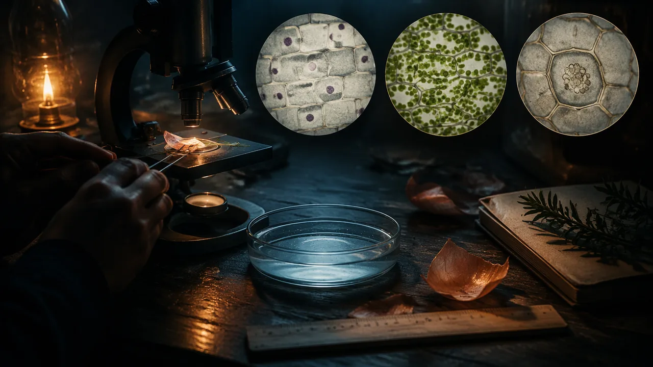
Why start with cells? Every function you studied in the launch, from sweating to pumping blood, is carried out by cells. Cell structures are matched to their jobs, and one structure matters for every cell: the plasma membrane, which controls what enters and leaves.
Cells possess specialized structures like the plasma membrane and cell wall that regulate their interaction with the environment, especially the movement of water, which is vital for survival.
📖 Read for this lesson
The reading opens over the lesson; close it to come back to exactly where you were. The checks below draw on it.
The thin, flexible boundary of every cell, made of a lipid bilayer with proteins, which controls what enters and leaves.
Before we look, what makes the plasma membrane such a critical structure for every cell to function?
A 10x eyepiece and a 40x objective give a total magnification of x. If the FOV at 40x is 4.5 mm, the FOV at that magnification is mm.
Plant and animal cells
The word ‘cell’ was first used by Robert Hooke in 1665 to describe the microscopic, box-like structures he observed in cork, which reminded him of the small rooms, or ‘cells,’ in a monastery.
All eukaryotic cells have a plasma membrane, cytoplasm, a nucleus holding DNA, ribosomes that build proteins, and mitochondria that release energy from food. Plant cells add three structures you will see today: a rigid cell wall of cellulose outside the membrane, a large central vacuole filled with water, and, in green tissue, chloroplasts. Onion bulb cells have no chloroplasts because the bulb grows underground; the Elodea leaf does.
A rigid layer outside the plasma membrane of plant cells (made of cellulose) that gives support and prevents bursting.
Which structure did you see in Elodea but not in onion bulb cells, and why?
Using the microscope
Total magnification is the eyepiece magnification times the objective magnification: a 10x eyepiece with a 40x objective gives 400x. The field of view (FOV), the diameter of the circle you see, shrinks as magnification rises, in inverse proportion. If the FOV is 4.5 mm at 40x, then at 400x (ten times the magnification) it is 4.5 ÷ 10 = 0.45 mm, or 450 µm. To estimate a cell’s length, count how many cells fit across the field. If 1.5 onion cells span the 400x field end to end, each is about 450 ÷ 1.5 = 300 µm long. Always measure the low-power FOV with a clear ruler first, because microscopes differ.
The diameter of the circle visible through a microscope; it decreases as magnification increases.
Plant cells always keep their rigid shape because of their strong cell wall.
While the cell wall provides rigid support, the living contents of a plant cell, including the plasma membrane and vacuole, can shrink or swell within the wall as water moves by osmosis, as seen in plasmolysis.
A cube-shaped cell with side 3 µm has a larger surface-area-to-volume ratio than one with side 1 µm.
Why are cells small?
A cell exchanges materials through its surface, but its needs grow with its volume. For a cube of side s, surface area is 6s2 and volume is s3, so the ratio is 6/s. A 1-unit cube has a ratio of 6; a 2-unit cube has 3; a 3-unit cube has only 2. As a cell grows, each unit of volume has less membrane to serve it. That is one reason large organisms are made of many small cells rather than a few huge ones.
Simulation & tools
Model osmosis at the membrane
Open the Membrane Transport sim. (1) Add about 50 sodium ions to the outside only and no channels. Record whether any cross. (2) Add two sodium leak channels and watch for one minute. Record which way the ions move and when the movement appears to stop. (3) Write two sentences: why does salt need a channel to cross, and how does that explain why water, not salt, moved in your Elodea cells? No internet? Use the section ‘The membrane as a gatekeeper’ in this lesson’s reading and answer the same questions.
Open in a new tab ↗ · PhET Interactive Simulations, University of Colorado Boulder · CC BY 4.0
Osmosis
The membrane lets water cross freely but blocks most dissolved salt. Water moves by osmosis from where it is more concentrated (fewer dissolved particles) to where it is less concentrated (more dissolved particles). Put a plant cell in salty water and water leaves the vacuole; the membrane pulls away from the rigid wall. This is plasmolysis, and it is why lettuce wilts under salad dressing and why a potato strip left in salt water turns limp and loses mass, which you will measure at home. In fresh water, water enters, the vacuole swells and pushes against the wall, making the cell firm (turgid). Animal cells have no wall, so in very dilute water they can swell and burst, which is one reason the body tightly regulates the salt and water in blood.
The movement of water across a membrane from a region of lower solute concentration to a region of higher solute concentration.
Shrinking of a plant cell’s contents away from the cell wall as water leaves by osmosis.
Connecting to the phenomenon
A worker who sweats heavily loses both water and salt. Drinking plain water replaces the water but not the salt; drinking nothing concentrates the body’s fluids. Either way, the concentration outside cells changes, and cells gain or lose water by osmosis. Homeostasis includes keeping that concentration steady.
Lab: Onion and Elodea cells on a virtual microscope, and what salt water does to plant tissue
Question. What structures can we see in plant cells, how big are they, and how does placing them in salt water change them?
Materials
- This lesson’s reading: the scaled micrograph plate (four 400x fields, each 450 µm across)
- Pencil, paper and a clear ruler
- One raw potato and a table knife or vegetable peeler (an adult can do the cutting)
- Two clear cups or glasses, table salt, tap water, a teaspoon and a measuring cup
- A kitchen scale, if your home has one (otherwise the ruler and a bend test are enough)
- A timer or phone clock, and paper towels
- Have a parent or guardian cut the potato, or cut it yourself only with their permission, on a cutting board with the blade moving away from your fingers.
- Do not eat the potato or taste any solution; pour the salt water down the sink, throw the potato away and wash your hands at the end.
- Wipe up spills at once so no one slips.
Procedure
- Open the micrograph plate in this lesson’s reading. Each circle is one 400x field of view, 450 µm across: the field of a microscope whose 40x field measures 4.5 mm. Record 4.5 mm as the FOV at 40x, and show the calculation that gives the FOV at 400x.
- Field 1 is onion bulb skin stained with iodine. Draw three or four cells and label the cell wall, nucleus and cytoplasm. Count how many cells fit end to end across the field through its center, counting a partial cell as the fraction you can see.
- Field 2 is an Elodea leaf in tap water. Draw it, labeling the chloroplasts and the cell wall, and count the cells along one row of the field.
- Fields 3 and 4 are the same Elodea cells after five minutes in salt solution and five minutes after flushing with tap water. Draw the same cells from each field and describe what moved and what stayed in place.
- Now test the same idea on plant tissue you can hold. Cut six potato strips about 1 cm by 1 cm by 6 cm. Measure each strip’s length to the nearest millimeter and, if you have a kitchen scale, weigh three strips together for each cup.
- Pour 100 mL (a little under half a cup) of tap water into one cup. Stir 1 teaspoon of table salt into 100 mL of tap water in the other. Put three strips in each cup and leave them for 60 minutes.
- Take the strips out, blot them dry and measure length and mass again. Hold each strip by one end and record whether it stays stiff or bends. Calculate the percentage change in mass (or length) for each cup. Optional: move the salt-water strips into fresh tap water for another 60 minutes and record whether they stiffen again.
Data
| Measurement | Value |
|---|---|
| FOV at 40x (mm) | |
| FOV at 400x (mm, calculated) | |
| Onion cells across the field (field 1) | |
| Estimated onion cell length (µm) | |
| Elodea cells along one row (field 2) | |
| Estimated Elodea cell length (µm) | |
| Elodea in tap water (field 2): description | |
| Elodea after 5 min in salt water (field 3): description | |
| Elodea back in tap water (field 4): description |
| Potato strips | Mass before (g) | Mass after (g) | Mean length before (mm) | Mean length after (mm) | % change | Stiff or bendy? |
|---|---|---|---|---|---|---|
| Tap water | ||||||
| Salt water |
Analysis
- Show your field-of-view calculation and your estimates of onion and Elodea cell length. Count the onion cells along two other lines across field 1, compare the three estimates, and suggest why they differ.
- Claim-evidence-reasoning: what does salt water do to plant cells? Use fields 2, 3 and 4 as cell-level evidence and your potato measurements as tissue-level evidence, with osmosis as your reasoning.
- Which structure kept the Elodea cells from bursting when they returned to tap water, and why did the tap-water potato strips turn stiff instead of bursting? What would happen to a cell without that structure?
- Name two sources of error in estimating cell size from a field of view and two in the potato test, and say how you would reduce each.
If you cannot do this lab in person: The main route above needs no microscope, and once the reading is open it needs no internet. Two other routes earn the same full credit and produce the same drawings, the same size calculation and the same claim-evidence-reasoning paragraph. Option A — your own microscope. If your home has a compound microscope or a clip-on phone microscope lens, peel the thin inner skin from an onion layer, lay it flat in a drop of water on a slide, stain it with a drop of diluted food coloring (not iodine), add a cover slip, and use your own cells in place of field 1, measuring your own 40x field with the clear ruler. Handle slides and cover slips by the edges and have an adult clear up any broken glass. Use fields 2, 3 and 4 of the plate for the Elodea drawings. Option C — the root-tip record in this lesson’s second reading. ‘An Onion Root Tip at 400x’ is an illustrative record of three stained fields from the growing tip of an onion root: 200 cells sorted by phase of the cell cycle, with the counting method stated, and 22 cells lying end to end across a 450 µm field. Draw and label three cells from the plate, then take your numbers from the root-tip record: express one root-tip cell as a fraction of the field width and convert it to micrometers, work out the percentage of the 200 cells in each phase and check that your five percentages sum to 100, and write two sentences comparing root-tip cells with onion-skin cells in size and in what they are doing. For the plasmolysis paragraph use fields 2, 3 and 4 of the plate. No potato or no scale? Use the illustrative potato results under ‘Plasmolysis you can hold’ in this lesson’s first reading as your tissue-level evidence. Optional, for any route: the PhET Membrane Transport simulation linked on this lesson’s sim card can be run as a measured experiment — count the sodium ions on each side at 0, 30 and 60 seconds with no channels, then with two leak channels — as extra evidence for why water, and not salt, crossed the Elodea membrane. It is a tool, not a source; nothing in the lab depends on it. Cite the reading you used.
Practice
Work these on paper or in your notebook, then open Check your answer. Aim for all of Fluency and Application; try at least one Challenge.
Printable version: this unit’s practice workbook (PDF)
Fluency
Build speed and accuracy with the core skill.
- A microscope has a 10x eyepiece. Find the total magnification with the 4x, 10x and 40x objectives.
Check your answer
Answer: 40x, 100x and 400xTotal = eyepiece × objective. - The field of view at 40x is 4.5 mm. What is it at 100x?
Check your answer
Answer: 1.8 mmFOV is inversely proportional to magnification: 4.5 × 40/100 = 1.8 mm. - Convert 0.45 mm to micrometers.
Check your answer
Answer: 450 µm1 mm = 1,000 µm. - Find the surface-area-to-volume ratio of a cube-shaped cell with side 4 µm.
Check your answer
Answer: 1.5 per µm (96 µm2 ÷ 64 µm3)SA = 6 × 42 = 96; V = 43 = 64; ratio = 6/s = 1.5.
Application
Use the skill in context. Show your reasoning.
- At 400x the field of view is 450 µm. Three onion cells fit end to end across it. Estimate the length of one cell.
Check your answer
Answer: About 150 µm450 ÷ 3 = 150 µm. - Five Elodea cells fit across the same 450 µm field. Estimate one cell’s length and compare it with the onion cell.
Check your answer
Answer: About 90 µm; the onion cell (150 µm) is about 1.7 times longer.450 ÷ 5 = 90 µm; 150 ÷ 90 ≈ 1.7. - An Elodea leaf is placed in a salt solution much saltier than its cell sap. Predict what happens to the vacuole and the membrane, and explain in terms of water movement.
Check your answer
Answer: The vacuole shrinks and the membrane pulls away from the wall (plasmolysis), because water moves by osmosis out of the cell toward the higher solute concentration.Water crosses the membrane freely; salt mostly does not, so water leaves the cell. - Three potato strips weighed 20 g together before 60 minutes in salt water and 17 g after; three strips in tap water went from 20 g to 21 g (illustrative results). Find each percentage change in mass and explain the difference.
Check your answer
Answer: Salt water: −15%; tap water: +5%. Water left the cells in salt water and entered them in tap water, by osmosis.(17 − 20) ÷ 20 = −0.15; (21 − 20) ÷ 20 = +0.05. Water moves toward the higher solute concentration: out of the cells in salt water, into them in tap water, where the cell walls keep them from bursting.
Challenge
Stretch problems. Expect to think before you write.
- Compare a flat cell measuring 1 × 10 × 10 µm with a cube of the same volume. Find each surface-area-to-volume ratio and explain why many cells that exchange materials quickly are flat.
Check your answer
Answer: Flat cell: SA = 240 µm2, V = 100 µm3, ratio 2.4. Cube of the same volume: side ≈ 4.64 µm, ratio ≈ 1.29. The flat shape nearly doubles the surface per unit volume, speeding exchange.SA = 2(1×10 + 1×10 + 10×10) = 240. Cube side = 1001/3 ≈ 4.64; ratio = 6/4.64 ≈ 1.29.
Review
Keep earlier skills sharp.
- Convert 100 kPa to atm.
Check your answer
Answer: 0.9869 atm
Road salt spread on Missouri highways in winter washes into roadside soil, where it can pull water out of plant root cells by osmosis; that is one reason shrubs and grass along salted roads often brown by spring.
How does understanding osmosis in plant cells help us explain the importance of homeostasis in our own bodies?
How confident are you that you can identify cell structures in a micrograph, estimate cell size from the field of view, and explain plasmolysis with osmosis?
The Shift
- Plant cells have a rigid cell wall and a plasma membrane.
- Microscopes allow us to visualize cells and estimate their size.
- Osmosis explains how cells gain or lose water.
Inside a Plant Cell: Microscope Fields, Membranes and Osmosis at Home
SLS reading for RLP Biology A, drawing on the NCI SEER training module on cell structure and the NHGRI Talking Glossary; the micrograph plate and the potato results are illustrative; retrieved November 5, 2026 · SLS original
What every cell shares, and what plant cells add
Your body and an onion look nothing alike, but their cells are built on the same plan. Every eukaryotic cell has a plasma membrane around it, a watery cytoplasm inside, a nucleus that holds the DNA, ribosomes that assemble proteins, and mitochondria that release energy from food. Plant cells add three structures you can see with an ordinary light microscope, or on the scaled plate later in this reading:
| Structure | Job | Onion bulb skin | Elodea leaf |
|---|---|---|---|
| Cell wall (cellulose) | Rigid support outside the membrane; stops the cell bursting | Yes | Yes |
| Large central vacuole | Stores water and dissolved substances; presses outward on the wall | Yes | Yes |
| Chloroplasts | Photosynthesis | No | Yes |
| Nucleus | Holds DNA; controls the cell | Yes (easy to see with iodine) | Yes (often hidden by chloroplasts) |
Why no chloroplasts in onion? The bulb is a storage organ that grows underground, in the dark, so its cells have no use for photosynthesis. Structure matches function: the cell makes the parts its job requires. In Elodea you may see chloroplasts gliding around the edge of each cell; the cytoplasm is streaming, a sign the cell is alive.
The membrane as a gatekeeper
The plasma membrane is a double layer of lipid molecules studded with proteins. Its oily middle lets small uncharged molecules such as oxygen and carbon dioxide slip through, and water also crosses readily. Charged particles, such as the sodium and chloride ions of dissolved table salt, are repelled by the oily layer. They cross only through channel proteins, tunnels that open a path for particular ions. In the PhET Membrane Transport simulation you can see this directly: sodium ions added to one side stay there until you add channels, then move through from the side where they are more concentrated to the side where they are less concentrated, and net movement stops once the two sides are about equal. Because salt needs channels and water does not, when a cell meets salty water it is mostly water that moves.
Osmosis in plant and animal cells
Osmosis is the movement of water across a membrane toward the side with more dissolved particles (the higher solute concentration). Think of dissolved particles as “holding on” to water: water drifts toward where more particles are holding it.
- Salty surroundings (more solute outside): water leaves the vacuole, the cytoplasm shrinks, and the membrane peels away from the wall. This is plasmolysis. The wall keeps its shape, so you see a green blob pulled away inside a rigid box.
- Fresh water (less solute outside): water flows in, the vacuole swells and presses against the wall, and the cell becomes firm, or turgid. The wall stops it from bursting. That is why plasmolysed Elodea recovers when you flush it with tap water.
- Animal cells have no wall. In very dilute water a red blood cell can swell and burst; in very salty water it shrivels. This is why the body keeps the salt and water content of blood within narrow limits, and why a worker who sweats heavily and drinks nothing, or drinks huge amounts of plain water, can upset that balance.
Magnification and field of view
Total magnification = eyepiece × objective. With a 10x eyepiece, the 4x, 10x and 40x objectives give 40x, 100x and 400x. The field of view (FOV) is the diameter of the bright circle. It shrinks in inverse proportion to magnification: multiply the magnification by some factor and the FOV is divided by the same factor.
- Measure the FOV at low power with a clear ruler. Suppose it is 4.5 mm at 40x.
- At 100x (2.5 times the magnification): 4.5 × 40/100 = 1.8 mm.
- At 400x (10 times): 4.5 ÷ 10 = 0.45 mm. Since 1 mm = 1,000 µm, that is 450 µm.
Other conversions you will meet in practice sets: 1 kg = 1,000 g, 1 L = 1,000 mL, 1 cm3 = 1 mL, kelvin = °C + 273.15, and km/h ÷ 3.6 = m/s.
To estimate cell size, count how many cells fit end to end across the high-power field and divide. If 1.5 onion cells span 450 µm, each is 450 ÷ 1.5 = 300 µm long. If 3 cells fit, each is 150 µm; if 5 Elodea cells fit, each is 90 µm, and the 150 µm onion cell is 150 ÷ 90 ≈ 1.7 times longer. Estimates differ from one count to the next because cells really do vary, because partial cells can be counted differently, and because each microscope’s FOV is slightly different. You reduce the error by measuring your own low-power FOV, counting along several lines, and averaging.
An illustrative micrograph plate
You do not need a microscope for this lab: work from the four fields below. If you do have one at home, they also let you check your own slide. They are drawn rather than photographed, which is why they are labeled illustrative — but they are drawn to scale, and every measurement the lab asks for can be taken off them. Each circle is one 400x field of view, 450 µm across, which is what you get from a microscope whose 40x field measures 4.5 mm; the bar at the bottom is 100 µm at the same magnification. Field 1 is onion bulb epidermis stained with iodine. Fields 2, 3 and 4 are the same patch of an Elodea leaf: in tap water, after five minutes in salt solution, and five minutes after flushing with tap water.
How to use the plate. For the size estimate, count how many onion cells fit end to end across field 1, counting a partial cell as the fraction you can see, and divide 450 µm by that number; do the same along one row of field 2 for the Elodea cells, and compare the two. For the drawings, copy three or four cells from field 1 and label the cell wall, the nucleus and the cytoplasm, then copy cells from field 2 and label the cell wall and the chloroplasts — the small circles set around the inside of each cell. For the claim-evidence-reasoning paragraph, compare fields 2, 3 and 4 and look carefully at what moved: in field 3 the contents have pulled away from the walls, and the walls themselves are exactly where they were. Fill in the lab’s table as you go, entering 4.5 mm as the field of view at 40x.
Plasmolysis you can hold: a potato test
The plate shows osmosis one cell at a time. A raw potato shows the same thing in a whole tissue, using only a kitchen. Cut six strips about 1 cm by 1 cm by 6 cm. Put three in 100 mL of tap water and three in 100 mL of water with 1 teaspoon of table salt stirred in. After 60 minutes, blot them dry, weigh and measure them again, and try bending each one. The results below are illustrative, typical of this test; yours will differ, and that is fine.
| Cup | Mass of 3 strips before (g) | After 60 min (g) | Change in mass | Mean length before (mm) | After (mm) | Feel |
|---|---|---|---|---|---|---|
| Tap water | 20 | 21 | +5% | 60 | 61 | Stiff; snaps when bent |
| Salt water | 20 | 17 | −15% | 60 | 57 | Limp; bends without breaking |
Percentage change = (after − before) ÷ before × 100. For the salt-water strips: (17 − 20) ÷ 20 = −0.15, a 15% loss. For the tap-water strips: (21 − 20) ÷ 20 = +0.05, a 5% gain. In length the salt-water strips lost (57 − 60) ÷ 60 = −5%. The explanation is the one the plate shows: salt water outside the cells has the higher solute concentration, so water leaves each cell by osmosis, the vacuoles shrink and the strip goes limp. In tap water, water enters, the vacuoles press against the cell walls and the strip turns stiff, or turgid; the walls stop the cells from bursting. Mass is a better measure than length here, because the strip shrinks in all three directions at once.
Why cells stay small
Everything a cell takes in or gets rid of must cross its surface, but how much it needs depends on its volume. For a cube of side s, surface area = 6s2 and volume = s3, so the surface-area-to-volume ratio is 6/s.
| Side s (µm) | Surface area (µm2) | Volume (µm3) | SA : V (per µm) |
|---|---|---|---|
| 1 | 6 | 1 | 6 |
| 2 | 24 | 8 | 3 |
| 3 | 54 | 27 | 2 |
| 4 | 96 | 64 | 1.5 |
As the cube grows, the ratio falls: each cubic micrometer of cell is served by less membrane. A big cell would starve for supplies at its center. Shape helps too. A flat cell 1 × 10 × 10 µm has surface area 2(1×10 + 1×10 + 10×10) = 240 µm2 and volume 100 µm3, a ratio of 2.4. A cube with the same volume has side 1001/3 ≈ 4.64 µm and a ratio of 6 ÷ 4.64 ≈ 1.29. Flattening nearly doubles the surface per unit volume, which is why cells that exchange materials quickly, such as those lining the lungs, are thin and flat.
Writing up plasmolysis
A claim-evidence-reasoning paragraph might read: Claim: salt water made water leave the Elodea cells. Evidence: after five minutes in salt solution, the green contents had shrunk away from the walls in most cells; after flushing with tap water, they filled the cells again; at the tissue scale, potato strips in salt water lost 15% of their mass and went limp, while strips in tap water gained 5% and turned stiff. Reasoning: the membrane lets water cross but blocks most salt, so water moved by osmosis toward the higher solute concentration outside; the rigid wall kept the cell’s outline and prevented bursting when water flowed back in.
Sources: NCI SEER Training: Structure of the Cell (training.seer.cancer.gov); NHGRI Talking Glossary: Cell Membrane (genome.gov); NHGRI Talking Glossary: Nucleus (genome.gov); NHGRI Talking Glossary: Mitochondria (genome.gov)
An Onion Root Tip at 400x: Cell Counts by Phase
SLS illustrative record for Biology A (n = 200 cells in three fields, method stated in the reading), drawing on the NHGRI Talking Glossary and MedlinePlus Genetics on cell division for the phase definitions · SLS original
Where an onion makes new cells
The onion skin you drew in this lesson is finished tissue: large, flat storage cells that will never divide again. To see cells being made, look at the other end of the plant. Behind the very tip of every root is a meristem, a few millimeters of small, boxy, unspecialized cells that divide over and over. At 400x the same field that shows four or five onion-skin cells shows more than twenty root-tip cells side by side, so a root tip gives you a second, very different data set for cell size, and it lets you count something the skin never shows: cells caught in the act of dividing.
How this record was made
This is an illustrative record prepared by SLS Learning as a stand-in for a slide you would make yourself; the numbers are typical of a healthy onion root tip but were not taken from any published data set. An onion bulb was set over a jar of water for five days until the roots were about 3 cm long. The terminal 2 mm of one root was cut off, fixed in household vinegar for ten minutes, softened in warm dilute hydrochloric acid, stained with toluidine blue, and squashed flat under a cover slip so that the cells lay in a single layer. It was viewed at 400x with a field of view 450 µm across. Three non-overlapping fields were chosen within 1 mm of the tip. Every cell whose nucleus lay entirely inside the field was assigned to one phase using the rules below; cells cut by the field edge were not counted. The three fields hold 200 cells in total (n = 200).
| Phase | Rule used to assign a cell |
|---|---|
| Interphase | Nucleus is a smooth, evenly stained circle; no separate chromosomes can be seen |
| Prophase | Nucleus looks grainy or threadlike as the chromosomes condense; nuclear outline fading |
| Metaphase | Chromosomes gathered in one dense band across the middle of the cell |
| Anaphase | Two groups of chromosomes pulling apart toward opposite ends, a clear gap between them |
| Telophase | Two dense clusters at opposite ends, with a new wall (the cell plate) starting to form between them |
The counts
| Field | Interphase | Prophase | Metaphase | Anaphase | Telophase | Cells in field |
|---|---|---|---|---|---|---|
| Field 1 | 55 | 7 | 2 | 1 | 2 | 67 |
| Field 2 | 54 | 6 | 3 | 1 | 2 | 66 |
| Field 3 | 55 | 7 | 2 | 2 | 1 | 67 |
| Total | 164 | 20 | 7 | 4 | 5 | 200 |
Check the row totals before you use the table: 55 + 7 + 2 + 1 + 2 = 67, and the three fields sum to 200. A count you have not checked is not evidence yet.
Turning counts into percentages
A percentage is a fraction of the total scaled to 100. For interphase: 164 ÷ 200 = 0.820, and 0.820 × 100 = 82.0%. Do the same for the other four phases, then add all five percentages; they must come to 100.0. The four dividing phases together (prophase through telophase) are 36 of the 200 cells, and that fraction, the mitotic index, is the usual single-number measure of how actively a tissue is dividing. In the onion skin from the same bulb the mitotic index is zero: no cell in the plate is dividing.
Notice that the three fields do not agree exactly. Field 2 has three metaphase cells and field 3 has two anaphase cells where the others have one. That is sampling variation, not a mistake, and it is why the record pools three fields instead of reporting one. If you were counting your own slide, more fields would give a steadier percentage.
Cell size from the same field
Along the diameter of field 1, from edge to edge through the center, 22 meristem cells lay end to end. One cell is therefore 1/22 of the field width. The field is 450 µm across, so one root-tip cell is about 450 ÷ 22 = 20.5 µm long, roughly a fiftieth of a millimeter. Use the same arithmetic on any micrograph: field width divided by the number of cells across it. If a picture carries no scale bar, that field width is the one measurement you would still need before the fraction can become micrometers.
Compare this with the onion-skin cells you measured in Option A or on the plate in this lesson’s first reading. Skin cells run several times longer than meristem cells because they have stopped dividing and have swelled with water stored in a large central vacuole; meristem cells are small because each one divides again before it has time to grow much. Size and activity go together, and the two records from one onion show both ends of that story.
Sources: NHGRI Talking Glossary: Mitosis (genome.gov); MedlinePlus Genetics: How do cells divide? (medlineplus.gov)