3.19 First Test: Magnification and Image Clarity
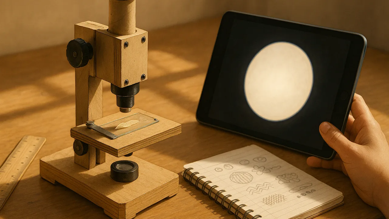
plan and run a fair test of our prototype’s magnification and image clarity using onion skin and elodea, and record the data.
- I can describe the data I will collect to show if living and nonliving things have cells.
- I can describe how I will look for parts of living things that are not made of cells.
- I can compare the presence and types of cells in different organisms, including those made of one cell or many cells.
- I can collect and record data about the cellular makeup of living organisms.
- I can identify the tools I use to observe cells at different magnifications and explain why different tools are needed to see cells at different scales.
Phase 4: Build
Warm-Up ~5 min
Do Now. Your lens makes a ruler 30 times larger. Is that enough to see onion cells? What else besides magnification makes a picture useful?
As magnification goes up, what usually happens to the area of the slide you can see?
Instruction ~10 min
Two measures describe image quality. Magnification is how much larger the image is. Clarity is how sharp the details are. A picture can be large and still blurry. We need both, because our criteria ask for at least 100× and a nucleus and cell wall clear enough to label.
A fair test. Every team photographs the same specimens: onion skin stained with iodine and an elodea leaf. Keep the light source, the distance and the specimens the same. Change only the station design. Score clarity with a class scale: 1 = no cells visible, 2 = cell outlines only, 3 = cell walls clear, 4 = walls and nuclei clear.
Worked example 1: field of view and cell size (sample data). Malik’s photo shows 2 mm of ruler across the screen. Eight onion cells fit end to end across the same view. Each cell is about 2 mm ÷ 8 = 0.25 mm long. That is about a quarter of a millimeter, which explains why we need a lens.
Worked example 2: different tools for different scales. A hand lens at about 10× shows that an onion layer is a thin sheet. A clip-on lens at about 30× shows the outlines of cells like bricks in a wall. A school microscope at 100× to 400× shows the nucleus inside each cell. A school microscope’s total magnification is the eyepiece times the objective, so 10× × 40× = 400×.
| Specimen | Design | Magnification (×) | Clarity score (1–4) | Parts we could see | Cells across the view |
|---|---|---|---|---|---|
| Onion + iodine | |||||
| Elodea leaf | |||||
| Paper scrap (nonliving) |
Guided Exercises ~15 min
Prepare one slide at a time, photograph it, then answer the next block.
Independent Practice ~10 min
Analyze your team’s results.
Copy your data table. Which specimen gave your best clarity score, and what parts could you see?
Does your version 1 meet the 100× criterion and show a clear nucleus? Use your magnification and clarity score as evidence and name one change for version 2.
Closure ~5 min
Good data, scientists. Next class: pixels, and why image size matters when you send a picture.
Exit ticket: explain why a scientist might need a hand lens, a clip-on lens AND a microscope to study one onion.
How confident are you that you can I can collect and record data about the cells of living things.
Teacher notes
Materials: onion, iodine solution in dropper bottles, elodea, slides, cover slips, droppers, paper towels, goggles, gloves, rulers, every team’s version 1, one school microscope for comparison.
Setup: demonstrate a wet mount once. Onion epidermis is easiest when peeled from the inner face of a layer. Elodea nuclei are hard to see; that is a useful result, not a failure. Cell sizes here are sample data.
Listen for: “more zoom is always better”; treating the paper fibers as cells.
Support / stretch: provide photo examples of each clarity score. Stretch: estimate how many onion cells would fit across 1 cm.